Surface Energy Gradients for Characterizing Cell-Material
Interactions
Introduction
Surface energy is a fundamental material property
that often affects biological interaction. This response may
be through a direct cell-material interaction, but more often
seems to be an indirect effect where surface energy dictates
protein adsorption which subsequently dictates cell response.
Thus, we have developed rapid methods for characterizing cell
response to variations in surface energy in order to gain a
better understanding of the relationships between surface energy,
protein adsorption and cell behavior.
Experimental Approach
An automated stage was used to move silanized
glass slides beneath a UV lamp such that the ends of the slide
are exposed to the light for varying amounts of time. UV exposure
causes oxidation on the surface of the slide such that a longer
exposure results in a more hydrophilic surface. Gradients that
range in water contact angles from 30? to 90? from end to end
can be created on a single slide and cell response to these
gradients has been examined. The development of these methods
for creating surface energy gradients provides us with a unique
tool which can be used to probe the fundamental correlations
between cell response and the surface energy of a material.
Results
Contact angle (water in air) of 15 gradient specimens.
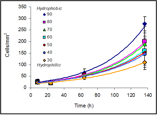
Cells (MC3T3-E1 osteoblasts) proliferated faster on hydrophobic
areas than on hydrophilic areas. Plot labels correspond to contact
angle.
Cell proliferation was directly proportional to surface energy.
Exponential factors from the fitted curves (from previous figure
above) plotted against contact angle produced a straight line.
Future Activities
Surface energy gradients could potentially be used for quality
control screening of cell stocks intended for human implantation.
Cell behavior on the gradients could be established and used
as a benchmark. The behavior of different batches of cells could
then be evaluated on the gradients as an indicator that they
have not transformed, mutated or lost their phenotype.
Publications
PAPER: Kennedy SB, Mei Y, Gross R, Washburn NR, Amis EJ.
(2005) Cell response on surface energy gradients. Biomaterials,
in preparation.
POSTER: Kennedy SB, Mei Y, Gross R, Washburn NR, Amis EJ.
(2003) Quantifying cell response to materials through population
analyses enabled by high-throughput techniques. Society for
Biomaterials 29th Annual Meeting, Reno, NV.
POSTER: Simon Jr CG, Kennedy SB, Amis EJ, Eidelman N, Washburn
NR. “Gradient Libraries for Combinatorial and High-Throughput
Investigations of Polymeric Biomaterials”, 7th World
Biomaterials Congress, Australia, 2004.
POSTER: Simon Jr CG, Kennedy SB, Amis EJ, Eidelman N, Washburn
NR. “High-throughput Methods for Biomaterials Development”,
NIST Combinatorial Methods Center 4th Annual Meeting, Gaithersburg,
MD, 2003.
POSTER: Simon Jr CG, Kennedy SB, Amis EJ, Eidelman N, Washburn
NR. “High-throughput Methods for Biomaterials Development”,
Symposium on Metrology and Standards for Cell Signaling, NIST,
Gaithersburg, MD, 2003.
POSTER: Simon Jr CG, Kennedy SB, Amis EJ, Eidelman N, “Washburn
NR. High-throughput Methods for Biomaterials Development”,
RESBIO Kickoff Even, Rutgers University, NJ 2003.
NIST Contributors:
Scott B. Kennedy
Ying Mei
Eric J. Amis
Newell R. Washburn
Collaborators:
Richard Gross
(Polytechnic University)
Biomaterials Group
Polymers Division
Materials Science and Engineering Laboratory