High-throughput Method for Characterizing Cell Response
to Polymer Crystallinity
Introduction
Surface topology can strongly influence the performance
of tissue engineered medical products. Crystalline polymers
used in biomedical applications, such as poly(e-caprolactone)
and poly(L-lactic acid), can have either a rough or a smooth
surface depending on how they are processed. When they are crystallized,
the surface becomes roughened but when they are kept amorphous
their surface remains smooth. Thus, we have used gradient technology
to develop a high-throughput method for studying cell response
to the surface roughness that results from polymer crystallinity.
Experimental Approach
Solutions of poly(L-lactic acid) (PLLA) were
spread onto glass substrates with a home-built flow-coater to
yield thin films of PLLA that were smooth and amorphous. The
films were placed on a temperature gradient stage such that
one end was held below the Tg at room temperature and the other
end was heated above the Tg to 100° C. This produced gradients
in crystallinity along the PLLA films where the room temperature-ends
remained amorphous and smooth while the 100° C-ends became
crystalline and roughened. The morphology of the gradients was
characterized with atomic force microscopy and cell response
on the films was assessed.
Results
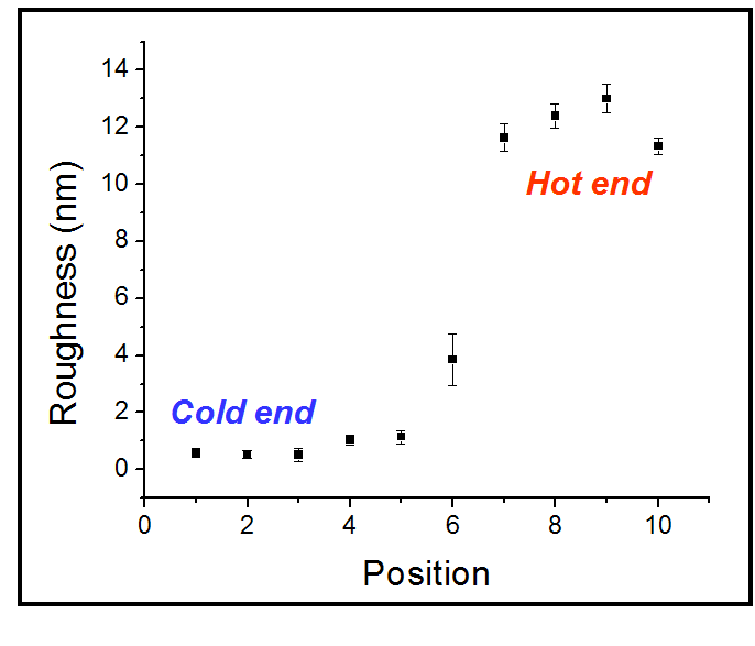
AFM was used to determine surface roughness (RMS) of a poly(L-lactide)
film annealed on a temperature gradient. The hot end became
rougher as spherulites began to form.
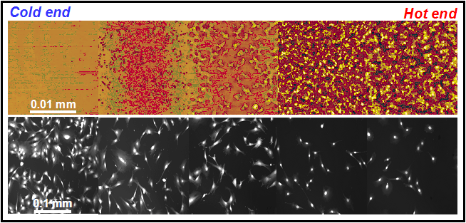
Upper images: A poly(L-lactide) film was annealed on a temperature
gradient and atomic force microscopy revealed that the hot end
became rougher as spherulites began to form. Lower images: Cells
(MC3T3-E1 osteoblasts) cultured (5d) on the gradient proliferated
faster on the smooth areas as determined by automated fluorescence
microscopy.
Left: Cell counting using automated fluorescence microscopy
confirmed that cell proliferation (5d, green) was enhanced on
the smooth end of the crystallinity gradients. Cell adhesion
after 1d was essentially equal across the gradient (yellow).
Right: A plot of cell number versus surface roughness showed
that the critical roughness for which a statistically significant
reduction in proliferation occurred was 4 ± 1 nm.
Future Activities
Polymer crystallinity gradients could potentially be used
for quality control screening of cell stocks intended for human
implantation. Cell behavior on the gradients could be established
and used as a benchmark. The behavior of different batches of
cells could then be evaluated on the gradients as an indicator
that they have not transformed, mutated or lost their phenotype.
Publications
PAPER: Washburn NR, Yamada KM, Simon Jr CG, Kennedy SB, Amis
EJ (2004) High-throughput investigation of osteoblast response
to crystalline polymers: influence of nanometer-scale roughness
on proliferation. Biomaterials 25, 1215-1224.
POSTER: Simon Jr CG, Kennedy SB, Amis EJ, Eidelman N, Washburn
NR. “Gradient Libraries for Combinatorial and High-Throughput
Investigations of Polymeric Biomaterials”, 7th World Biomaterials
Congress, Australia, 2004.
POSTER: Simon Jr CG, Kennedy SB, Amis EJ, Eidelman N, Washburn
NR. “High-throughput Methods for Biomaterials Development”,
NIST Combinatorial Methods Center 4th Annual Meeting, Gaithersburg,
MD, 2003.
POSTER: Simon Jr CG, Kennedy SB, Amis EJ, Eidelman N, Washburn
NR. “High-throughput Methods for Biomaterials Development”,
Symposium on Metrology and Standards for Cell Signaling, NIST,
Gaithersburg, MD, 2003.
POSTER: Simon Jr CG, Kennedy SB, Amis EJ, Eidelman N, “Washburn
NR. High-throughput Methods for Biomaterials Development”,
RESBIO Kickoff Even, Rutgers University, NJ 2003.
POSTER: Washburn NR, Kennedy SB, Simon Jr CG, Yamada KM, Amis
EJ. “High-Throughput Investigation of Cell Proliferation
on Crystalline Polymers”, Society for Biomaterials 29th
Annual Meeting, Reno, NV, 2003.
POSTER: Washburn NR, Kennedy SE, Sehgal A, Simon Jr CG, Amis
EJ. “High-throughput Investigations of Cell-Material Interactions”,
Gordon Research Conference on Signal Transduction by Engineered
Extracellular Matrices, New London, CT, 2002.
NIST Contributors:
Newell R. Washburn
Carl G. Simon, Jr.
Scott B. Kennedy
Eric J. Amis
Kathryn L. Beers
Collaborators:
Kenneth M. Yamada
(NIH/NIDCR )
Biomaterials Group
Polymers Division
Materials Science and Engineering Laboratory