Types of Holes in the Heart
Atrial Septal Defect
An atrial septal defect (ASD) is a hole in the part
of the septum that separates the atria (upper chambers of the heart). This
heart defect allows oxygen-rich blood from the left atrium to flow across the
atrial septum into the right atrium instead of flowing down to the left
ventricle as it should. This is inefficient because oxygen-rich blood gets
pumped back to the lungs, where it has just been, instead of going to the body.
Cross-Section of a Normal Heart and
a Heart With Atrial Septal Defect
Figure A shows the normal structure
and blood flow in the interior of the heart. Figure B shows a heart with an
atrial septal defect, which allows oxygen-rich blood from the left atrium to
mix with oxygen-poor blood from the right atrium.
An ASD can be small or large. Small ASDs allow only
a little blood to flow from one atrium to the other. Small ASDs don't affect
the way the heart works and therefore don't need any special treatment. Many
small ASDs close on their own as the heart grows during childhood.
Medium to large ASDs allow more blood to leak from
one atrium to the other, and they are less likely to close on their own. Most
children with ASDs have no symptoms, even if they have large ASDs.
There are three major types of ASD:
- Secundum. This defect is in the middle of the
atrial septum. It’s the most common form of ASD. About 8 out of every 10
babies born with ASD have secundum defects. At least half of all secundum ASDs
close on their own. This is less likely if the defect is large.
- Primum. This defect is in the lower part of the
atrial septum. It often occurs along with abnormalities in the heart valves
that connect the upper and lower heart chambers. Primum defects aren’t
very common. This type of defect doesn’t close on its own.
- Sinus venosus. This defect is in the upper part
of the atrial septum, near where a large vein (the superior vena cava) brings
oxygen-poor blood from the upper body to the right atrium. Sinus venosus is a
rare defect. Sinus venosus defects don’t close on their own.
Long-Term Effects of Atrial Septal Defects That
Aren’t Repaired
Over time, the extra blood flow to the right side of
the heart and the lungs may cause problems for a heart that has an ASD.
Usually, most of these problems don’t show up until adulthood, often
around age 30 or later. They are rare in infants and children. These possible
problems include:
- Right heart failure. The right side of the heart
has to work harder to pump extra blood to the lungs. Over time, the heart may
become tired from this extra work and not pump efficiently.
-
Arrhythmias (irregular heartbeats). Extra blood flowing into the right
atrium through an ASD can cause the atrium to stretch and enlarge. Over time,
this can lead to problems with the heart’s rhythm. When this occurs, an
arrhythmia can develop, with signs or symptoms such as palpitations (a feeling
that your heart has skipped a beat or is beating too hard) or a rapid
heartbeat.
- Stroke. Usually, the lungs filter out small clots
that can form on the right side of the heart. Sometimes a blood clot formed on
the right side of the heart can pass through an ASD to the left side and be
pumped out to the body. A clot like this can travel to an artery in the brain,
blocking blood flow through it and causing a stroke. This doesn’t occur in
childhood.
-
Pulmonary arterial hypertension (PAH). PAH is high blood pressure in the
arteries in the lungs. Over time, high blood pressure in the lungs can damage
the arteries and the small blood vessels in the lungs. They thicken and become
stiff, making it harder for blood to flow through them.
These problems develop over many years and
don’t occur in children. They also are rare in adults because most ASDs
either close on their own or are repaired in early childhood.
Ventricular Septal Defect
A ventricular septal defect (VSD) is a hole in the
part of the septum that separates the ventricles, the lower chambers of the
heart. The hole allows oxygen-rich blood to flow from the left ventricle across
the heart into the right ventricle instead of flowing up into the aorta and out
to the body as it should.
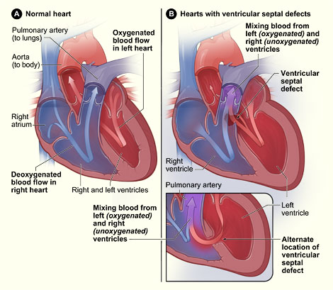
Cross-Section of a Normal Heart and
a Heart With Ventricular Septal Defect
Figure A shows the normal structure
and blood flow in the interior of the heart. Figure B shows two common
locations for a ventricular septal defect. The defect allows oxygen-rich blood
from the left ventricle to mix with oxygen-poor blood in the right ventricle.
An infant born with a VSD may have a single hole or
more than one hole in the wall that separates the two ventricles. The defect
also may occur by itself or with other congenital heart defects.
Doctors classify VSDs based on the:
- Size of the defect.
- Location of the defect.
- Number of defects.
- Presence or absence of a ventricular septal
aneurysm—a thin flap of tissue on the septum. This tissue is harmless and
can help a VSD close on its own.
VSDs can be small or large. A small VSD doesn’t
cause problems and may often close on its own. Because small VSDs allow only a
small amount of blood to flow between the ventricles, they’re sometimes
called restrictive VSDs. Small VSDs don’t cause any symptoms.
Medium VSDs are less likely than small defects to
close on their own. They may require surgery to close and may cause symptoms
during infancy and childhood.
Large VSDs allow a large amount of blood to flow
from the left ventricle to the right ventricle and are sometimes called
nonrestrictive VSDs. A large VSD is less likely to close completely on its own,
but it may get smaller over time. Large VSDs often cause symptoms in infants
and children, and surgery is usually needed to close them.
VSDs are found in different parts of the septum.
- Membranous VSDs are located near the heart
valves. They can close at any time.
- Muscular VSDs are found in the lower part of the
septum. They’re surrounded by muscle, and most close on their own during
early childhood.
- Inlet VSDs are located close to where blood
enters the ventricles. They’re less common than membranous and muscular
VSDs.
- Outlet VSDs are found in the part of the
ventricle where the blood leaves the heart. This is the rarest type of VSD.
Long-Term Effects of Large Ventricular Septal
Defects That Aren’t Repaired
A moderate to large VSD can cause:
-
Heart
failure. Infants with large VSDs may develop heart failure because the left
side of the heart pumps blood into the right ventricle in addition to its
normal work of pumping blood to the body. The increased workload on the heart
also increases the heart rate and the body’s demand for energy.
- Growth failure, especially in infancy. A baby may
not be able to eat enough to keep up with his or her body’s increased
energy demands. As a result, the baby may lose weight or fail to grow and
develop normally.
- Arrhythmias (irregular heartbeats). The extra
blood flowing through the heart can cause areas of the heart to stretch and
enlarge. This can disturb the normal electrical activity of the heart, leading
to fast and irregular heart rhythms.
- PAH. The high pressure and high volume of extra
blood pumped through a large VSD into the lungs can cause scarring of the
delicate arteries in the lungs. Today, PAH rarely develops because most large
VSDs are repaired in infancy.
|