How Is Coronary Angioplasty Done?
Before coronary angioplasty is done, your doctor
will need to know whether your coronary arteries are blocked. If one or more of
your arteries are blocked, your doctor will need to know where and how severe
the blockages are.
To find out, your doctor will do an
angiogram
and take an x-ray picture of your arteries. During an angiogram, a small tube
called a catheter with a balloon at the end is put into a large blood vessel in
the groin (upper thigh) or arm. The catheter is then threaded to the coronary
arteries. A small amount of dye is injected into the coronary arteries and an
x-ray picture is taken.
This picture will show any blockages, how many, and
where they're located. Once your doctor has this information, the angioplasty
can proceed. Your doctor will blow up (inflate) the balloon in the blockage and
push the plaque outward against the artery wall. This opens the artery more and
improves blood flow.
Coronary Balloon Angioplasty
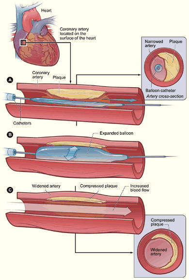
The illustration shows a
cross-section of a coronary artery with plaque buildup. The coronary artery is
located on the surface of the heart. Figure A shows the deflated balloon
catheter inserted into the narrowed coronary artery. In figure B, the balloon
is inflated, compressing the plaque and restoring the size of the artery.
Figure C shows the widened artery.
A small mesh tube called a
stent
is usually placed in the newly widened part of the artery. The stent holds up
the artery and lowers the risk of the artery renarrowing. Stents are made of
metal mesh and look like small springs.
Some stents, called drug-eluting stents, are coated
with medicines that are slowly and continuously released into the artery. These
medicines help prevent the artery from becoming blocked again from scar tissue
that grows around the stent.
Stent Placement

The illustration shows
the placement of a stent in a coronary artery with plaque buildup. Figure A
shows the deflated balloon catheter and closed stent inserted into the narrowed
coronary artery. The inset image on figure A shows a cross-section of the
artery with the inserted balloon catheter and closed stent. In figure B, the
balloon is inflated, expanding the stent and compressing the plaque to restore
the size of the artery. Figure C shows the stent-widened artery. The inset
image on figure C shows a cross-section of the compressed plaque and
stent-widened artery.
In some cases, plaque is removed during
angioplasty. In a procedure called atherectomy (ath-er-EK-toe-me), a catheter
with a rotating shaver on its tip is inserted into the artery to cut away
plaque. Lasers also are used to dissolve or break up the plaque. These
procedures are now rarely done because angioplasty gives better results for
most patients.
|
