What Does Cardiac CT Show?
Many x rays are taken while you’re in the
computed tomography (CT) scanner. Each picture that the machine takes shows a
small slice of the heart. A computer can put the pictures together to make a
large picture of the whole heart. This picture shows the inside of the heart
and the structures that surround the heart.
Cardiac CT
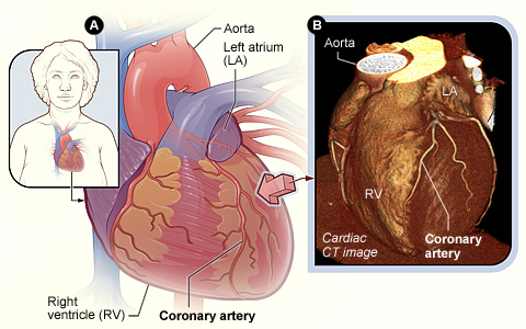
Figure A shows the position of the
heart in the body. The arrow shows the point of view of the cardiac CT image.
Figure B is a cardiac CT image showing the coronary arteries on the surface of
the heart. This is a picture of the whole heart, put together by the computer.
Cardiac CT is a common test for finding and
evaluating:
- Problems in the heart. Iodine-based dye used with
a cardiac CT scan can show pictures of the coronary arteries. The coronary
arteries are blood vessels on the surface of the heart. If these blood vessels
are narrowed or blocked, you may have chest pain or a
heart
attack. The CT scan also can find problems with heart function and heart
valves.
- Problems with the aorta. The aorta is the main
artery that carries oxygen-rich blood from the heart to the body. Cardiac CT
can detect two serious problems in the aorta:
-
Aneurysms, which are diseased areas of a weak blood vessel wall that bulge
out. Aneurysms can be life threatening because they can burst.
- Dissections, which can occur when the layers of
the aortic artery wall peel away from each other. This condition can cause pain
and also may be life threatening.
- Blood clots in the lungs. A cardiac CT scan also
may be used to find a
pulmonary
embolism, a serious but treatable condition. A pulmonary embolism is a
sudden blockage in a lung artery, usually due to a blood clot that traveled to
the lung from the leg.
- Pericardial disease. This is a disease that
occurs in the pericardium, a sac around your heart.
Because the heart is in motion, a fast type of CT
scanner, called multidetector computed tomography (MDCT), is used to take
high-quality pictures of the heart.
Another type of CT scanner, called electron-beam
computed tomography (EBCT), is used to detect calcium in the coronary arteries.
Calcium in the coronary arteries may be an early sign of
coronary
artery disease (CAD). |