
Chapter 5: Beyond Drug Design
This booklet has focused on drug design as the most immediate medical application of structural biology. But detailed studies of protein structure have value and potential far beyond the confines of the pharmaceutical industry. At its root, such research teaches us about the fundamental nature of biological molecules. The examples below provide a tiny glimpse into areas in which structural biology has, and continues to, shed light.
Muscle Contraction
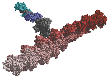
Click for larger image.
With every move you make, from a sigh to a sprint, thick ropes of myosin muscle proteins slide across rods of actin proteins in your cells. These proteins also pinch cells in two during cell division and enable cells to move and change shape—a process critical both to the formation of different tissues during embryonic development and to the spread of cancer. Detailed structures are available for both myosin and actin.
Transcription and Translation
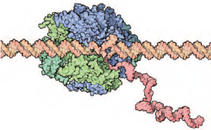
Click for larger image.
Cells use DNA instructions to make proteins. Dozens of molecules (mostly proteins) cling together and separate at carefully choreographed times to accomplish this task. The structures of many of these molecules are known and have provided a better understanding of transcription and translation.
A key example is RNA polymerase, an enzyme that reads DNA and synthesizes a complementary strand of RNA. This enzyme is a molecular machine composed of a dozen different small proteins. In 2001, Roger Kornberg, a crystallographer at Stanford University, determined the structure of RNA polymerase in action. This crystal structure suggested a role for each of RNA polymerase's proteins. Kornberg was awarded the 2006 Nobel Prize in Chemistry for this work.
Photosynthesis
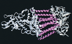
Click for larger image.
"Photosynthesis is the most important chemical reaction in the biosphere, as it is the prerequisite for all higher life on Earth," according to the Nobel Foundation, which awarded its 1988 Nobel Prize in chemistry to three researchers who determined the structure of a protein central to photosynthesis.
This protein, from a photosynthetic bacterium rather than from a plant, was the first X-ray crystallographic structure of a protein embedded in a membrane. The achievement was remarkable, because it is very difficult to dissolve membrane-bound proteins in water—an essential step in the crystallization process.
To borrow further from the Nobel Foundation: "[This] structural determination…has considerable chemical importance far beyond the field of photosynthesis. Many central biological functions in addition to photosynthesis…are associated with membrane-bound proteins. Examples are transport of chemical substances between cells, hormone action, and nerve impulses"—in other words, signal transduction.
Signal Transduction
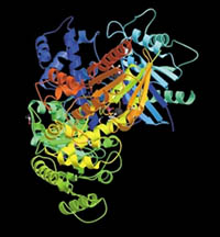
Click for larger image
Hundreds, if not thousands, of life processes require a biochemical signal to be transmitted into cells. These signals may be hormones, small molecules, or electrical impulses, and they may reach cells from the bloodstream or other cells.
Once signal molecules bind to receptor proteins on the outside surface of a cell, they initiate a cascade of reactions involving several other molecules inside the cell. Depending on the nature of the target cell and of the signaling molecule, this chain of reactions may trigger a nerve impulse, a change in cell metabolism, or the release of a hormone. Researchers have determined the structure of some molecules involved in common signal transduction pathways.
The receptor proteins that bind to the original signal molecule are often embedded in the cell's outer membrane so, like proteins involved in photosynthesis, they are difficult to crystallize.
Obtaining structures from receptor proteins not only teaches us more about the basics of signal transduction, it also brings us back to the pharmaceutical industry. At least 50 percent of the drugs on the market target receptor proteins—more than target any other type of molecule.
As this booklet shows, a powerful way to learn more about health, to fight disease, and to deepen our understanding of life processes is to study the details of biological molecules— the remarkable structures of life.
Got It?
Considering this booklet as a whole, how would you define structural biology?
What are the scientific goals of those in the field?
If you were a structural biologist, what proteins or systems would you study? Why?