|
Ophthalmological Disorders
In this series of paintings of the inside of the eye, the orange circle
represents the retina and the yellow circle represents the optic disc,
which is where the optic nerve that connects the eye to the brain leaves
the retina.
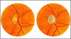
Painting 1: Glaucomatous Optic Disc
In the normal optic disc on the left, the number of optic nerve fibers
is normal. The cup within the optic nerve is small and the vessels are
near the center of the disc. In the glaucomatous optic disc on the right,
increased pressure within the eye has caused the disappearance of a large
number of optic nerve fibers. Therefore, the cup has enlarged and the
disc vessels have curved along the cup's contour.
Painting 01
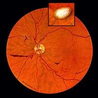
Painting 2: Retina Uveitis Hemorrhages
In 1958, Howard Bartner created his first painting at the National Institutes
of Health: this view of the retina of a patient with uveitis, an inflammation
of the uvea (iris, ciliary body, and choroid), the middle layer of the
eye. As a result, the retinal vessel walls are weakened and are bleeding.
The inset depicts a scar in the retina; the retina and choroid have disappeared
and the whiteness of the sclera (the outer layer of the eye) can be seen.
Painting 02
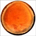
Painting 3: Detached Retina
This painting, completed in 1967, shows an entire retina. The optic disc
is the small yellow circle near the center of the orange retina. The billowing
of the bottom segment of the retina indicates that this segment of the
retina has become detached from the firm connective tissue that encloses
the eye. This causes visual loss in the area.
Painting 03
Back To Top | Photography
Credits
Related Links
National Eye Institute
Glaucoma Research Foundation
|