|
Jeffrey J. Neil, M.D., Ph.D.
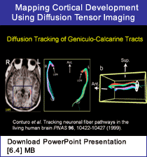
 SUMMARY: Diffusion tensor imaging (DTI) is a method of magnetic resonance imaging that measures water diffusion across several tissue axes, thus providing information regarding microstructure. It is useful for brain imaging because water displacements are not the same throughout the brain. In white matter, displacements are smaller, or more restricted than and perpendicular to myelinated fibers as they create physical boundaries that slow the diffusion of water molecules. This restricted diffusion is "anisotropic." Brain injury of any kind instantly causes a reduction in water displacement and diffusion coefficients making DTI an excellent diagnostic tool for early detection of brain injury and the occurrence of stroke. In this presentation, Dr. Jeffrey Neil detailed the specific principles of DTI, and gave examples of its use by several researchers and clinicians in studies of brain injury, normal brain morphology, and developing human and baboon brains.
In brain development studies, cortical grey matter in adult human subjects shows low values for diffusion anisotropy. However, early in development, the cortex shows very strong anisotropy. This has also has been observed in DTI studies of the developing baboon brain. These observations may represent more prominent laminar brain organization and less radial organization during brain development; however, more work needs to be done in human infants at various stages of development to confirm this. DTI is even useful with fixed tissue. Although there is less diffusion in fixed tissue, anisotropy and directional correlations are identical to those in the living human brain.
Neil concluded that the advent of DTI will allow the development of white matter fiber tracking algorithms and provide a rapid, noninvasive exam to better study and document white matter changes in cognitive processes. The use of DTI to measure and monitor cognitive and motor performances opens a new vista in the field of neuroimaging for early intervention and remediation of neural dysfunction.
|