
|
Contents:
Section I: Introduction to the Brain
Click on the images to see a larger version [file sizes range from 150K
to 260K], once you have viewed the image, use the "Back" button on your
browser to return to this page

|
1: Introduction
Introduce the purpose of your presentation. Indicate that you will
explain how the brain basically works and how and where drugs such
as heroin and cocaine work in the brain. Tell your audience that
you will discuss the concept of "reward" which is the property that
is characteristic of many addictive drugs.
|
|
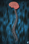
2: The brain and spinal cord
The central nervous system is composed of both the brain and the
spinal cord. Describe the brain as a functional unit; it is made
up of billions of nerve cells (neurons) that communicate with each
other using electrical and chemical signals.
|
|
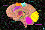
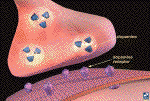
3: Brain regions and neuronal pathways
Certain parts of the brain govern specific functions. Point to areas
such as the sensory (orange), motor (blue) and visual cortex (yellow)
to highlight their specific functions. Point to the cerebellum (pink)
for coordination and to the hippocampus (green) for memory. Indicate
that nerve cells or neurons connect one area to another via pathways
to send and integrate information. The distances that neurons extend
can be short or long. For example, point to the reward pathway (orange).
Explain that this pathway is activated when a person receives positive
reinforcement for certain behaviors ("reward"). Indicate that you
will explain how this happens when a person takes an addictive drug.
As another example, point to the thalamus (magenta). This structure
receives information about pain coming from the body (magenta line
within the spinal cord), and passes the information up to the cortex.
Tell the audience that you can look at this in more detail.
|

|
4: Pathway for sensation of pain and reaction to pain
This is a long pathway, in which neurons make connections in both
the brain and the spinal cord. Explain what happens when one slams
a door on one's finger. First, nerve endings in the finger sense
the injury to the finger (sensory neurons) and they send impulses
along axons to the spinal cord (magenta pathway). Point to each
part of the pathway as you explain the flow of information. The
incoming axons form a synapse with neurons that project up to the
brain. The neurons that travel up the spinal cord then form synapses
with neurons in the thalamus, which is a part of the midbrain (magenta
circle). The thalamus organizes this information and sends it to
the sensory cortex (blue), which interprets the information as pain
and directs the nearby motor cortex (orange) to send information
back to the thalamus (green pathway). Again, the thalamus organizes
this incoming information and sends signals down the spinal cord,
which direct motor neurons to the finger and other parts of the
body to react to the pain (e.g., shaking the finger or screaming
"ouch!").
|
|
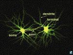
5: Neuronal structure
Indicate that these pathways are made up of neurons. This image
contains real neurons from the thalamus. They have been filled with
a fluorescent dye and viewed through a microscope. Describe the
anatomy of a neuron: point to the cell body (soma), dendrites, and
axon (marked with text). At the end of the axon is the terminal,
which makes a connection with another neuron. [Note: the axon has
been drawn in for clarity, but actually, the axons of these neurons
travel to the cerebral cortex.]
|
|
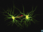
6: Impulse flow
Explain the normal direction of the flow of information (electrical
and chemical). An electrical impulse (the action potential) travels
down the axon toward the terminal. Point to the terminal. The terminal
makes a connection with the dendrite of neighboring neuron, where
it passes on chemical information. The area of connection is called
the synapse. Although the synapse between a terminal and a dendrite
(shown here) is quite typical, other types of synapses exist as
well. For example, a synapse can occur between a terminal and a soma
or axon.
|
|
7: The synapse and synaptic neurotransmission
Describe the synapse and the process of chemical neurotransmission.
As an electrical impulse arrives at the terminal, it triggers vesicles
containing a neurotransmitter, such as dopamine (in blue), to move
toward the terminal membrane. The vesicles fuse with the terminal
membrane to release their contents (in this case, dopamine). Once
inside the synaptic cleft (the space between the two neurons) the
dopamine can bind to specific proteins called dopamine receptors
(in pink) on the membrane of a neighboring neuron. This is illustrated
in more detail on the next image.
|
|
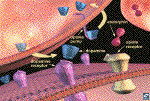
8: Dopamine neurotransmission and modulation by endogenous
opiates
Using the close-up of a synapse, continue using dopamine for your
example of synaptic function. Explain that it is synthesized in
the nerve terminal and packaged in vesicles. Reiterate the steps
in neurotransmission. Show how the vesicle fuses with the membrane
and releases dopamine. The dopamine molecules can then bind to a
dopamine receptor (in pink). After the dopamine binds, it comes
off the receptor and is removed from the synaptic cleft by uptake
pumps (also proteins) that reside on the terminal (arrows show the
direction of movement). This process is important because it ensures
that not too much dopamine remains in the synaptic cleft at any
one time. Also point out that there are neighboring neurons that
release another compound called a neuromodulator. Neuromodulators
help to enhance or inhibit neurotransmission that is controlled
by neurotransmitters such as dopamine. In this case, the neuromodulator
is an "endorphin" (in red). Endorphins bind to opiate receptors
(in yellow) which can reside on the post-synaptic cell (shown here)
or, in some cases, on the terminals of other neurons (this is not
shown so it must be pointed out). The endorphins are destroyed by
enzymes rather than removed by uptake pumps.
|
[Previous Section] [Next
Section]
|
|

Teacher Information
Here are some other NIDA-related sites which may be of interest.
Click on any of the links below to view those sites.
|
|


