What To Expect During Stress Testing
During all types of stress testing, a technician
will always be with you to closely monitor your health status.
Before you start the “stress” part of a
stress test, a technician will put small sticky patches called electrodes on
the skin of your chest, arms, and legs. To help an electrode stick to the skin,
the technician may have to shave a patch of hair where the electrode will be
attached.
The electrodes are connected to a machine that
records the electrical activity of your heart. This recording, which is called
an EKG
(electrocardiogram), shows how fast your heart is beating and the heart’s
rhythm (steady or irregular). The machine also records the strength and timing
of electrical signals as they pass through each part of your heart.
The technician will put a blood pressure cuff on
your arm to monitor your blood pressure during the stress test. (The cuff will
feel tight on your arm when it expands every few minutes.) In addition, you may
be asked to breathe into a special tube so the gases you breathe out can be
monitored.
After these preparations, you will exercise on a
treadmill or stationary bicycle. If such exercise poses a problem for you, you
may instead turn a crank with your arms. During the test, the exercise level
will get harder. But you can stop whenever you feel the exercise is too much
for you.
Stress Testing
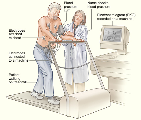
The illustration shows a patient
having a stress test. Electrodes are attached to the patient’s chest and
connected to an EKG (electrocardiogram) machine. The EKG records the
heart’s electrical activity. A blood pressure cuff is used to record the
patient’s blood pressure while he walks on a treadmill.
If you can’t exercise, a technician will inject
a medicine into a vein in your arm or hand. This medicine will increase the
flow of blood through the coronary arteries and/or make your heart beat faster,
as would exercise. This results in your heart working harder, so the stress
test can be performed. The medicine may make you flushed and anxious, but the
effects disappear as soon as the test is over. The medicine may also give you a
headache.
While you’re exercising or receiving medicine
to make your heart work harder, the technician will ask you frequently how
you’re feeling. You should tell him or her if you feel chest pain,
shortness of breath, or dizzy. The exercise or medicine infusion will continue
until you reach a target heart rate, or until you:
- Feel moderate to severe chest pain
- Get too out of breath to continue
- Develop abnormally high or low blood pressure or
an arrhythmia
(an abnormal heartbeat)
- Become dizzy
The technician will continue to monitor your heart
functions and blood pressure for a short time after you stop exercising or stop
receiving the stress-creating medicine. The “stress” part of a stress
test (when you’re exercising or given a medicine that makes your heart
work hard) usually lasts only about 15 minutes or less. But there is
preparation time before the test and monitoring time afterward. Both extend the
total test time to about an hour for a standard stress test, and up to 3 hours
or more for some imaging stress tests.
Exercise Stress Echocardiogram Test
For an exercise stress echocardiogram test, the
technician will take pictures of your heart using
echocardiography before you exercise and after you finish. A
sonographer (a person who specializes in using ultrasound techniques) will
apply a gel to your chest and then will briefly put a wand-like device (called
a transducer) against your chest and move it around. The transducer sends and
receives high-pitched sounds that you usually can’t hear. The echoes from
the sound waves are converted into moving pictures of your heart on a
screen.
You may be asked to lie on your side on an examining
table for this test. Some stress echocardiogram tests also use a dye to improve
imaging. This dye is injected into your bloodstream while the test occurs.
Sestamibi Stress Test or Other Imaging Stress Test
Involving Radioactive Dye
For a sestamibi or other imaging stress test that
uses a radioactive dye, the technician will inject a small amount of the dye
(such as sestamibi) into your bloodstream via a needle placed in a vein of your
arm or hand. You’re usually given the dye about a half-hour before you
start exercising or are given a medicine that makes your heart work hard. The
amount of radiation in the dye is safe and not a danger to you or those around
you. However, if you’re pregnant, you shouldn’t have this test
because of risks it might pose to your unborn child.
Pictures will be taken of your heart at least two
times—when it’s at rest and when it’s working its hardest. For
such imaging, you will lie down on a table and a special camera or scanner that
can see the dye in your bloodstream will take pictures of your heart. Some
pictures may not be taken until you lie quietly for a few hours after
exercising or receiving the stress-creating medicine. Some patients may even be
asked to return in a day or so for more pictures to be taken.
Magnetic Resonance Imaging Stress Test
A magnetic resonance imaging (MRI) stress test may
use a medicine rather than exercise to get your heart to work harder. But some
facilities have you exercise on a specially made bicycle or treadmill that
allows you to exercise while lying on your back. For this test, you will be put
inside a tunnel-like MRI machine that takes pictures of your heart when
it’s working hard and when your body is at rest. |