
 Computed axial tomography, commonly known as CAT scanning, was introduced in 1972. During a CAT scan, a large coil of x-ray tubes rotates around the patient's body, taking x-rays from all angles. A computer integrates all of these x-rays into a single, three-dimensional image on a television screen. The data can be saved on the computer. Allan Cormack, a high energy physicist at Tufts University, shared the 1979 Nobel Prize in Physiology and Medicine for his key work in developing the methods for CAT scanners. At the time of development, these methods were widely regarded as the most significant advance in medical radiography since the 1895 discovery of x-rays.
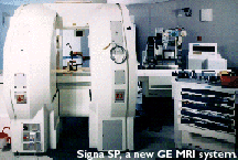
Magnetic Resonance Imaging (MRI) is used to localize brain activity during sensory or cognitive stimulation of the subject. Images of the subject's brain at rest and then during the performance of an intellectual task are compared to probe cognitive processes.
Sir Peter Mansfield and Paul Lauterbur won the 2003 Nobel Prize in Physiology or Medicine "for their discoveries concerning magnetic resonance imaging". CAT and MRI scanners have revolutionized diagnosis of disorders of soft tissues, especially disorders of the head and brain. Most shock-trama units or major neurological clinics have one of these machines on-site or at their immediate disposal. The sophisticated mathematical techniques used to reconstruct the images of organs and tissues that doctors see with these amazing diagnostic instruments originated in particle detection methods developed by high-energy physicists. For more information about CAT and MRI scanners, see the database report The Ultimate Structure of Matter: The High Energy Physics Program from the 1950s Through the 1980s.
|


 |
Information
Bridge • Energy
Citations Database • E-print
Network • R&D
Accomplishments
About OSTI Science.gov • USA.gov • USAJOBS • Grants • Regulations.gov |
|---|

