What To Expect During Carotid Ultrasound
Carotid ultrasound is usually done in a
doctor’s office or hospital. The test is painless and usually doesn’t
take more than 30 minutes.
The ultrasound machine includes a computer, a video
screen, and a transducer, which is a hand-held device that sends and receives
ultrasound waves into and from the body.
You will lie down on your back on an exam table for
the test. Your technician or doctor will put a gel on your neck where your
carotid arteries are located. This gel helps the ultrasound waves reach the
arteries better. Your technician or doctor will put the transducer against
different spots on your neck and move it back and forth.
The transducer gives off ultrasound waves and
detects their echoes after they bounce off the artery walls and blood cells.
Ultrasound waves can’t be heard by the human ear.
A computer uses the echoes of the ultrasound waves
bouncing off the carotid arteries to create and record images of the insides of
the arteries (usually in black and white) and your blood flowing through them
(usually in color; this is the Doppler ultrasound). A video screen displays
these live images for your doctor to review.
Carotid Ultrasound
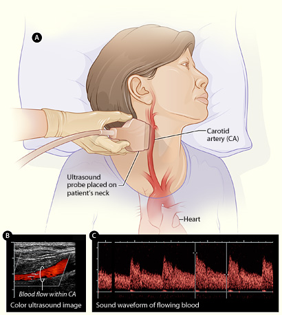
Figure A shows how the ultrasound
probe is placed over the carotid artery. Figure B is a color ultrasound image
showing blood flow (the red color in the image) in the carotid artery. Figure C
is a waveform image showing the sound of flowing blood in the carotid
artery. |