How Is Peripheral Arterial Disease Diagnosed?
Peripheral arterial disease (PAD) is diagnosed based
on general medical and family history, history of leg or heart problems,
personal risk factors, a physical exam, and test results. An accurate diagnosis
is critical, because people with PAD face a six to seven times higher risk of
heart disease or
stroke than the rest of the population. PAD is often diagnosed
after symptoms are reported. If you have PAD, your doctor also may want to look
for signs of
coronary
artery disease (CAD).
Specialists Involved
Mild PAD may be managed by a primary care doctor,
internist, or general practitioner. For more advanced PAD, a vascular
specialist (a doctor who specializes in treating blood vessel problems) may be
involved. A cardiologist (a doctor who specializes in heart diseases) also may
be involved in the care of patients with PAD.
Medical and Family History
Medical and family history is important in
diagnosing PAD. Your doctor may:
- Ask about your family history of cardiovascular
disease
- Review your medical history, including
high
blood pressure or
diabetes
- Ask about any symptoms, including any symptoms
that occur when walking or exercising
- Ask if you are currently or used to be a smoker
- Ask if you have any symptoms in the legs when
sitting, standing, walking, or climbing
- Review your diet
- Review your current medicines
Physical Exam
The physical exam may involve:
- Checking blood flow in your leg or foot to see if
the pulse is either weak or absent.
- Checking pulses in your leg arteries for an
abnormal whooshing sound called a bruit (broo-E). A bruit can be heard with a
stethoscope and may be a warning of a narrow or blocked section of an
artery.
- Checking for poor wound healing.
- Comparing blood pressure between your limbs to
see if blood pressure is lower in the affected limb.
- Checking hair, skin, and nails for any changes
that may indicate PAD.
Diagnostic Tests and Procedures
A simple test called an ankle-brachial index (ABI)
can be used to diagnose PAD. The ABI compares blood pressure in the ankle with
blood pressure in the arm to see how well blood is flowing. A normal ABI is 1.0
or greater (with a range of 0.90 to 1.30). The test takes about 10–15
minutes to measure both arms and both ankles. It can help the doctor find out
if PAD is affecting the legs, but it will not identify which blood vessels are
blocked. The ABI can be performed yearly if necessary to see if the disease is
getting worse.
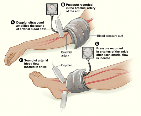
The illustration shows the
ankle-brachial index (ABI) test. The ABI gives the ratio of the systolic blood
pressure in the ankle to the systolic blood pressure in the brachial artery of
the arm.
A
Doppler ultrasound is a test that uses sound waves to tell
whether a blood vessel is open or blocked. This test uses a blood pressure cuff
and special device to measure blood flow in the veins and arteries in the arms
and legs. The Doppler ultrasound can help to determine the level and degree of
PAD.
A treadmill test will provide more information on
the severity of the symptoms and the level of exercise that provokes symptoms.
For this test, you will walk on a treadmill, which will help identify any
difficulties that you may have during normal walking.
A magnetic resonance angiogram (MRA) uses radio wave
energy to take pictures of blood vessels inside the body. MRA is a type of
magnetic resonance imaging (MRI) scan. An MRA can detect problems that may
cause reduced blood flow in the blood vessels. It can determine the location
and degree of blockage. A patient with a pacemaker, prosthetic joint, stent,
surgical clips, mechanical heart valve, or other metallic devices in his or her
body might not be eligible for an MRA depending on the type of metallic
device.
An arteriogram is a "road map" of the arteries used
to pinpoint the exact location of the blockage in a limb. An
x ray is taken after injecting dye through a needle or
catheter into an artery. When the dye is injected, the patient may feel mildly
flushed. The pictures from the x ray can determine the location, type, and
extent of the blockage. Some hospitals are using a newer method that uses tiny
ultrasound cameras to take pictures inside the blood vessel.
Blood tests may be done to check the patient's blood
sugar level to screen for diabetes. Blood tests also may be used to check the
patient's cholesterol levels.
|