U.S. Food & Drug Administration
Center for
Food
Safety &
Applied
Nutrition
Bacteriological
Analytical
Manual
Online
January 2001
|
|
Chapter 15
Bacillus
cereus Diarrheal Enterotoxin
Author
(Return to Table of Contents)

Bacillus cereus is an aerobic sporeformer that is commonly found in soil, on
vegetables, and in many raw and processed foods. Consumption of foods that
contain large numbers of B. cereus (106 or more/g) may result in food
poisoning, especially when foods are prepared and held for several hours
without adequate refrigeration before serving. Cooked meat and vegetables,
boiled or fried rice, vanilla sauce, custards, soups, and raw vegetable
sprouts have been incriminated in past outbreaks (1). Two types of illness
are attributed to the consumption of foods contaminated with B. cereus. The
first and better known is characterized by abdominal pain and diarrhea; it has
an incubation period of 4-16 h and symptoms that last for 12-24 h (4,5). The
second, which is characterized by an acute attack of nausea and vomiting that
occurs within 1-5 h after a meal; diarrhea is not a common feature in this
type of illness.
Although certain physiological and cultural characteristics are necessary for
identifying B. cereus (4), its enterotoxigenicity indicates whether a suspect
strain may be a public health hazard. Evidence shows that diarrheal toxin is a
distinct serological entity; in vitro methods that use specific antibodies
have been developed to detect the toxin in culture fluids. The evidence for
the emetic toxin, however, is still incomplete. This chapter presents a
method for the routine culturing of suspect Bacillus spp., using a semisolid
agar medium and a serological procedure (the microslide gel double diffusion
test) to identify the enterotoxin.
- Equipment and materials
- Test tubes, 25 x 100 and 20 x 150 mm
- Petri dishes, 15 x 100 and 20 x 150 mm, sterile
- Bottles, prescription, 4 oz
- Microscope slides, glass, pre-cleaned, 3 x 1 inch (7.62 x 2.54 cm)
- Pipets, sterile, 1, 5, and 10 ml, graduated
- Centrifuge tubes, 50 ml
- Sterile bent glass spreaders
- Electrical tape, 0.25 mm thick, 19.1 mm wide, available from
Scotch Brand, 3M Co., Electro-Products Division, St. Paul, MN
55011.
- Templates, plastic (Fig. 1)
- Silicone grease, high vacuum, available from Dow Corning Corp.,
Midland, MI 48640
- Sponges, synthetic
- Wooden applicator sticks
- Glass tubing, 7 mm, for capillary pipets and de-bubblers
- Pasteur pipets or disposable 30 or 40 µl
pipets, available from
Kensington Scientific Corp., 1165-67th St., Oakland, CA 94601, if
capillary pipets are not available.
- Staining jars (Coplin or Wheaton)
- Desk lamp
- Incubator, 35 ± 1°C
- Hot plate, electric
- Sterilizer (Arnold), flowing steam
- Blender and sterile blender jars (see Chapter 1)
- Centrifuge, high speed
- Timer, interval
- Media and reagents
- Brain heart infusion (BHI) broth (M24)
- Glucose, dextrose anhydrous
- Gel diffusion agar, 1.2% (R28)
- Nutrient agar slants (M112)
- Distilled water, sterile
- Phosphate-buffered dilution water (Butterfield's buffer) (R11)
- Normal (physiological) saline, sterile (antisera diluent) (R63)
- Thiazine Red R stain (R79)
- Slide preserving solution for stained slides, 1% acetic acid and
1% glycerol (R69)
- No. 1 McFarland standard (R42)
- Antisera and reference enterotoxins
- Preparation of materials and media
- BHIG, 0.1%. Adjust BHI broth containing 0.1% glucose to pH 7.4 and
dissolve by stirring. Distribute medium in 30 ml portions in 125
ml flasks and autoclave at 121°C for 10 min.
- No. 1 McFarland standard. Prepare turbidity standard No. 1 of
McFarland nephelometer scale (5). Mix 1 part 1% BaCl2 with 99
parts 1% H2SO4 in distilled water.
- 1.2% Gel diffusion agar for gel diffusion slides. Prepare fluid
base for agar in distilled water as follows: NaCl 0.85%; sodium
barbital 0.8%; merthiolate 1:10,000 (crystalline), available from
Eli Lilly and Co., Terre Haute, IN. Adjust pH to 7.4. Prepare
agar by adding 1.2% Noble special agar (Difco). Melt agar mixture
in Arnold sterilizer (steamer) and filter while hot, in steamer,
through 2 layers of filter paper; dispense in small portions
(15-25 ml) in 4 oz prescription bottles. (Remelting more than
twice may break down purified agar.)
- Thiazine Red R stain. Prepare 0.1% solution of Thiazine Red R
stain in 1.0% acetic acid.
- Preparation of slides. Wrap double layer of electrician's plastic
insulating tape around both sides of glass slide, leaving 2.0 cm
space in center. Apply tape as follows: Start a piece of tape
9.5-10 cm long about 0.5 cm from edge of undersurface of slide and
wrap tightly around slide twice. Wipe area between tapes with
cheesecloth soaked with 95% ethanol, and dry with dry cheesecloth.
Coat upper surface area between tapes with 0.2% agar in distilled
water as follows: Melt 0.2% bacteriological grade agar, and
maintain at 55°C or higher in screw-cap bottle. Hold slide over
beaker placed on hot plate adjusted to 65-85°C and pour or brush
0.2% agar over slide between 2 pieces of tape. Let excess agar
drain into beaker. Return agar collected in beaker to original
container for reuse. Wipe undersurface of slide. Place slide on
tray and dry in dust-free atmosphere (e.g., incubator). NOTE: If
slides are not clean, agar will roll off slide without coating it
uniformly.
- Preparation of slide assembly. Prepare plastic templates as
described by Casman et al. (2) (Fig. 1). Spread thin film of
silicone grease on side of template that will be placed next to
agar, i.e., the side with the smaller holes. Place about 0.4 ml
melted and cooled (55-60°C) 1.2% diffusion agar between tapes.
Immediately lay silicone-coated template on melted agar and edges
of bordering tapes. Place one edge of template on one of the
tapes and bring opposite edge to rest gently on the other tape.
Place slide in prepared petri dish (see C-7, below) soon after
agar solidifies and label slide with number, date, or other
information.
- Preparation of petri dishes for slide assemblies. Maintain
necessary high humidity by saturating 2 strips of synthetic sponge
(about 1/2 inch wide x 1/2 inch deep x 2-1/2 inches long) with distilled
water and placing them in each 20 x 150 mm petri dish. From 2 to 4
slide assemblies can be placed in each dish.
- Recovery of used slides and templates. Clean slides without
removing tape; rinse with tap water, brush to remove agar gel,
boil in detergent solution for 15-20 min, rinse about 5 min in hot
running water, and boil in distilled water. Place slides on end,
using test tube rack or equivalent, and place in incubator to dry.
If slides cannot be uniformly coated with hot 0.2% agar, they are
not clean enough and must be washed again. Avoid exposure to
excessive heat or plastic solvents when cleaning plastic
templates. Place templates in a pan and pour hot detergent
solution over them; let them soak 10-15 min. Use soft nylon brush
to remove residual silicone grease. Rinse sequentially with tap
water, distilled water, and 95% ethanol. Spread templates on
towel to dry.
- Directions for dissolving reagents used in slide gel. The reagents
are supplied as lyophilized preparations of enterotoxins and their
antisera. Rehydrate antisera in physiological saline. Rehydrate
reference enterotoxins in physiological saline containing 0.3%
proteose peptone, pH 7.0, or physiological saline containing 0.37%
dehydrated BHI broth, pH 7.0. These preparations should produce
faint but distinct reference lines in the slide gel diffusion
test. The lines may be enhanced (see E-3, below).
- Procedure for enumeration and selection of B. cereus colonies. For
examining food products, use procedures described for detecting B.
cereus (see Chapter 14). Test isolates for enterotoxigenicity as
described in E, below.
Production of enterotoxin. Of the methods described for the production
of enterotoxin, cultivation of B. cereus in BHIG (0.1% glucose, pH 7.4)
is simple and requires no special apparatus other than a shaker. Add
loopful of growth from nutrient agar slants to 3-5 ml sterile distilled
water or saline. Inoculate BHIG with 0.5 ml of this aqueous suspension,
which should contain about 300 million organisms/ml. Turbidity of
suspension should be equivalent to No. 1 on McFarland nephelometer
scale. Deliver suspension with sterile 1.0 ml pipet. Shake flasks at 3 ±
2°C at 84-125 cycles/ml for 12 h. Good surface growth is obtained
after 12 h of incubation. Transfer contents of flasks to 50 ml
centrifuge tube. Remove organisms by high speed centrifugation (10 min
at 32,800 x g). Examine supernatant for presence of enterotoxin by
filling depots in slide gel diffusion assembly, as directed in E, below.
- Slide gel diffusion test. To prepare record sheet, draw hole pattern of
template on record sheet, indicate contents of each well, and give each
pattern on record sheet a number to correspond with number on slide.
- Addition of reagents (Fig. 2). Place suitable dilution of
anti-enterotoxin (antiserum) in central well and place homologous
reference enterotoxin in upper peripheral well (if diamond pattern
is used); place material(s) under examination in well adjacent to
well containing reference enterotoxin(s). Use reference toxins and
antitoxins (antiserum), previously balanced, in concentrations
that give line of precipitation about halfway between their
respective wells. Adjust dilutions of reagents to give distinct
but faint lines of precipitation for maximum sensitivity. (See
C-9 for directions for dissolving reagents.) Prepare control slide
with only reference toxin and antitoxin.
Fig. 1. Microslide assembly with diagram for preparation and specifications
for plastic template.
Fill wells to convexity with reagents, using Pasteur pipet
(prepared by drawing out glass tubing of about 7 mm od) or
disposable 30 or 40 µl pipet. Remove bubbles from all wells by
probing with fine glass rod. Make rods by pulling glass tubing
very fine, as in making capillary pipets, breaking it into about
2-1/2 inch lengths, and melting ends in flame. It is best to fill
wells and remove bubbles against a dark background. Insert rods
into all wells to remove trapped air bubbles that may not be
visible. Let slides remain at room temperature in covered petri
dishes containing moist sponge strips for 48-72 h before
examination or for 24 h at 37°C.
- Reading the slide. Remove template by sliding it to one side. If
necessary, clean slide by dipping momentarily in water and wiping
bottom of slide; then stain as described below. Examine slide by
holding over source of light and against dark background.
Identify lines of precipitation through their coalescence with
reference line of precipitation (Fig. 3). If concentration of
enterotoxin in test material is excessive, formation of reference
line will be inhibited; test material must then be diluted and
retested. Figure 4, diagram A, shows typical precipitate line
inhibition caused by enterotoxin excess in test preparation
reactant arrangement in Fig. 2. Figure 5 shows typical line
formation. Figure 6 shows a diluted preparation. Occasionally,
atypical precipitate patterns that form may be difficult for
inexperienced analysts to interpret. One of the most common
atypical reactions is formation of lines not related to toxin but
caused by other antigens in test material (Fig. 7).
- Staining of slides. Enhance lines of precipitation by immersing
slide in Thiazine Red R strain for 5-10 min, and then examine.
Such enhancement is necessary when reagents have been adjusted to
give lines of precipitation that are only faintly visible. Use
staining procedure described by Crowle (3), modified slightly,
when slide is to be preserved. Rinse away any reactant liquid
remaining on slide by dipping slide momentarily in water and
immersing it for 10 min in each of the following baths: 0.1%
Thiazine Red R in 1% acetic acid; 1% acetic acid; and 1% acetic
acid containing 1% glycerol. Drain excess fluid from slide and
dry in 35°C incubator for storage as permanent record. After
prolonged storage, lines of precipitation may not be visible until
slide is immersed in water.

Fig. 2. Reagent arrangement for serologic identification of B.
cereus diarrheal antigen: 1) antiserum to B. cereus
antigen; 2) test preparation; 3) B. cereus toxin reference; 4) and 5) test preparations.
Fig. 3. Microslide gel diffusion test as toxin detection system; Antiserum to B. cereus
diarrheagenic antigen is in well 1; known reference enterotoxin is in well 3 to produce reference lines;
test preparations are in wells 2 and 4. Interpret reactions as follows: 1) No line development
between test preparations and antisera -- absence of B. cereus toxin; 2) coalescence
of test preparation line from well 4 with enterotoxin reference line -- presence of enterotoxin
in well 4; 3) coalescence of test preparation line from wells 2 and 4 with enterotoxin reference
-- presence of enterotoxin in wells 2 and 4.
Fig. 4. Effect of amount of B. cereus enterotoxin
in test preparation on development of reference line of precipitation. A, inhibition
(suppression) of reference line when 10 and 5 µg enterotoxin/ml, respectively, are
used. B-E, precipitate patterns when successively less enterotoxin (test preparation) is used. F, typical formation
of reference line of precipitation in slide test control system.
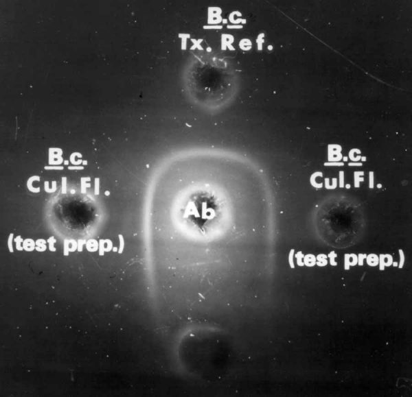
Fig. 5. Diarrheal antigen-antibody line of precipitation with microslide test.
Interpretation of reaction: B. cereus culture fluids (right, left, and
adjacent to reference toxin) contain diarrheagenic component, indicated by
lines of precipitation coalescing with reference line.
Fig. 6. Typical lines of precipitation of diluted B. cereus culture fluids,
using microslide test.
Fig. 7. B. cereus enterotoxin: Precipitate patterns in microslide gel
diffusion test demonstrate nonspecific (atypical) lines of precipitation
caused by other antigens reacting with nonenterotoxin antibodies. Test
preparations (wells 2 and 4) are negative for enterotoxin but produce
nonspecific lines of precipitation which intersect enterotoxin reference
lines of precipitation.
References
1. Bennett, R. W., and S. M. Harmon. 1988. Bacillus cereus food poisoning.
In: Laboratory Diagnosis of Infectious Diseases: Principles and
Practice, Vol. I, pp. 83-93. A. Balows, W. J. Hausler, Jr., M. Ohashi,
and A. Turano (eds). Springer-Verlag, New York.
2. Casman, E. P., R. W. Bennett, A. E. Dorsey, and J. E. Stone. 1969. The
micro-slide gel double diffusion test for the detection and assay of
staphylococcal enterotoxins. Health Lab. Sci. 6:185-198.
3. Crowle, A. J. 1958. A simplified micro double-diffusion agar precipitin
technique. J. Lab. Clin. Med. 52:784.
4. Lancette, G. A., and S. M. Harmon. 1980. Enumeration and confirmation of
Bacillus cereus in foods: collaborative study. J. Assoc. Off. Anal.
Chem. 63:581-586.
5. McFarland, J. 1907. The nephelometer: an instrument for estimating the
number of bacteria in suspensions used for calculating the opsonic index
and for vaccines. J. Am. Med. Assoc. 49:1176.
Hypertext Source: Bacteriological Analytical Manual,
8th Edition, Revision A, 1998. Chapter 15. Bacillus cereus Diarrheal Enterotoxin
*Author: Reginald W. Bennett
Top
B A M |
B A M Media |
B A M Reagents |
Bad Bug Book
Foods Home |
FDA Home |
Search/Subject Index |
Disclaimers & Privacy Policy
|
Accessibility/Help
Hypertext updated by kwg/cjm 2001-OCT-24