
National Wildlife Health Center
Multimedia
Search here for some of our available field imagery and videos.

Distribution of Chronic Wasting Disease in North America
Distribution of Chronic Wasting Disease in North America, updated December 17, 2020.

Histology panel from eastern gray squirrel
Photomicrographs from the lung of an eastern gray squirrel (Sciurus carolinensis) found dead in Wisconsin, U.S.A. (A) A bronchiole (arrow) contains numerous neutrophils. H&E stain. Inset: Bronchiolar epithelium is overlain by Gram-negative bacteria. Brown and Hopps stain. Bar = 20 µm. (B) Approximately 30% of the pulmonary parenchyma is replaced by dense
...
Necropsy photos from eastern gray squirrel
Photographs from an eastern gray squirrel (Sciurus carolinensis) found dead in Wisconsin, U.S.A. (A) The lung contained multifocal firm red areas (arrows) and multifocal areas of hemorrhage (arrowhead). (B) Firm areas are red to tan on cut section and airways contain mucoid red to tan fluid.

Photomicrographs from a desert cottontail found dead in Texas
Photomicrographs from a desert cottontail found dead in Texas, U.S.A. showing (A) panlobular hepatocellular dissociation and necrosis and (B) intra-alveolar edema fluid (asterisk) and foamy pulmonary macrophages (arrows) in the lung.

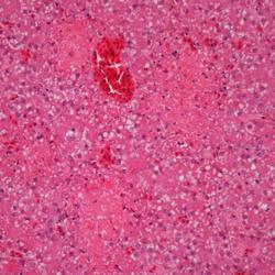
Photographs from organs of a mountain cottontail rabbit found dead
Photographs from an adult female mountain cottontail rabbit (Sylvilagus nuttallii) found dead in Montana, U.S.A. (A) Liver with random foci of necrosis (asterisk) characterized by accumulation of cellular detritus intermingled with fibrin and colonies of small coccoid bacteria (inset). H&E stain. (B) The spleen exhibits multifocal confluent areas of necrosis (
...
Photographs of intestine tissue from mallard ducks with trematodiasis
Photographs from two mallards (Anas platyrhynchos) found dead within a warm-water retention pond in Minnesota, US. (A) Caeca with mucosal roughening and caseous plaque formation. A larger trematode (arrow) is found within. (B) Dilated and thinned intestine containing watery intestinal content with pinpoint white material.

Photographs from two mallards (Anas platyrhynchos) with trematodiasis
Photographs from two mallards (Anas platyrhynchos) found dead within a warm-water retention pond in Minnesota, US. (A) Jejunum with superficial pseudomembranes and deep mucosal granulomatous inflammation (clear arrow). Small degenerated metazoan parasites are present rarely within the inflamed areas (black arrow). H&E stain. (B). Cecum with approximately 50 x
...
Photographs from a Common Eider (Somateria mollissima)
Photographs from a Common Eider (Somateria mollissima) from Massachusetts, USA. (A) There are scattered, small, pale foci in the liver. (B) Petechial and ecchymotic hemorrhages are present in the pancreas.
Photomicrographs from a Common Eider (Somateria mollissima)
Photomicrographs from a Common Eider (Somateria mollissima) from Massachusetts, USA. (A) Multiple pale foci of acute hepatic necrosis. (B) Multiple foci of acute pancreatic necrosis (arrow).

Images from necropsy of two foothill yellow-legged frogs (Rana boylii)
Two foothill yellow-legged frogs (Rana boylii) found dead in Santa Clara, California, USA. (A) One animal had pinpoint red foci on the ventral abdomen. (B) Another animal had a diffusely reddened kidney (arrow).

Photomicrographs from a foothill yellow-legged frog (Rana boylii)
Photomicrographs from a foothill yellow-legged frog (Rana boylii) found dead in Santa Clara, California, USA. (A) Small areas of epidermal necrosis with apoptotic keratinocytes and nuclear debris are multifocally present (arrow). (B) The liver shows randomly distributed, variably sized areas of coagulative necrosis (*). A few adjacent hepatocytes have
...
Photograph from a muskrat (Ondatra zibethicus) found dead in Ohio, USA
Photograph from a muskrat (Ondatra zibethicus) found dead in Ohio, USA. The liver contained disseminated pinpoint to 1-mm diameter white foci (arrows). Tyzzer's disease.