Image Library

The newsroom image library is home to the images journalists request most often. These high-resolution, public domain images are ready to print in your publication.

For images not available in this library, visit the Public Health Image Library (PHIL). We also recommend the National Library of Medicineexternal icon image library.

Viruses / Bacteria
Coronavirus
Coronavirus

PHIL ID #23354 | Download high-resolution image
Description:
Transmission electron microscopic image of an isolate from the first U.S. case of COVID-19, formerly known as 2019-nCoV. The spherical viral particles, colorized blue, contain cross-section through the viral genome, seen as black dots.

Coronavirus
Coronavirus

PHIL ID #23311 | Download high-resolution image
Description:
Transmission electron microscopic image of an isolate from the first U.S. case of COVID-19, formerly known as 2019-nCoV. The spherical extracellular viral particles contain cross-sections through the viral genome, seen as black dots.

Coronavirus
Coronavirus

PHIL ID #23311 | Download high-resolution image
Description:
This illustration, created at the Centers for Disease Control and Prevention (CDC), reveals ultrastructural morphology exhibited by coronaviruses. Note the spikes that adorn the outer surface of the virus, which impart the look of a corona surrounding the virion, when viewed electron microscopically. A novel coronavirus, named Severe Acute Respiratory Syndrome coronavirus 2 (SARS-CoV-2), was identified as the cause of an outbreak of respiratory illness first detected in Wuhan, China in 2019. The illness caused by this virus has been named coronavirus disease 2019 (COVID-19).

Coronavirus
Coronavirus

PHIL ID #23312 | Download high-resolution image
Description:
This illustration, created at the Centers for Disease Control and Prevention (CDC), reveals ultrastructural morphology exhibited by coronaviruses. Note the spikes that adorn the outer surface of the virus, which impart the look of a corona surrounding the virion, when viewed electron microscopically. A novel coronavirus, named Severe Acute Respiratory Syndrome coronavirus 2 (SARS-CoV-2), was identified as the cause of an outbreak of respiratory illness first detected in Wuhan, China in 2019. The illness caused by this virus has been named coronavirus disease 2019 (COVID-19).

Coronavirus
Coronavirus

PHIL ID #23313 | Download high-resolution image
Description:
This illustration, created at the Centers for Disease Control and Prevention (CDC), reveals ultrastructural morphology exhibited by coronaviruses. Note the spikes that adorn the outer surface of the virus, which impart the look of a corona surrounding the virion, when viewed electron microscopically. In this view, the protein particles E, S, and M, also located on the outer surface of the particle, have all been labeled as well. A novel coronavirus, named Severe Acute Respiratory Syndrome coronavirus 2 (SARS-CoV-2), was identified as the cause of an outbreak of respiratory illness first detected in Wuhan, China in 2019. The illness caused by this virus has been named coronavirus disease 2019 (COVID-19).

Escherichia coli (E. coli)
Coronavirus

PHIL ID #10042 | Download high-resolution image
Description:
Under a high magnification of 21674X, this digitally-colorized, scanning electron microscopic (SEM) image depicts a view of a dividing, Escherichia coli bacterium, clearly displaying the point at which the bacteria’s cell wall was splitting into two separate organisms. See PHIL 7137 for a black and white version of this image.

Listeria
Listeria

PHIL ID #21917 | Download high-resolution image
Description:
This illustration depicts a three-dimensional (3D), computer-generated image of a grouping of Listeria monocytogenes bacteria. The artistic recreation was based upon scanning electron microscopic (SEM) imagery.

Campylobacter
Campylobacter

PHIL ID #23238 | Download high-resolution image
Description:
This is a medical illustration of drug-resistant, Campylobacter sp. bacteria, presented in the Centers for Disease Control and Prevention (CDC) publication entitled, Antibiotic Resistance Threats in the United States, 2019 (AR Threats Report).

Measles
Measles

PHIL ID #21074 | Download high-resolution image
Description:
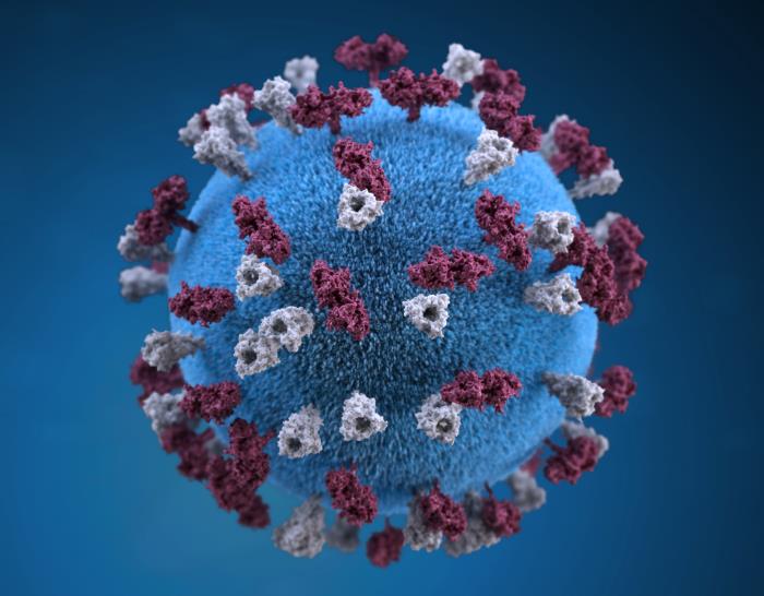
This illustration provided a 3D graphic representation of a spherical-shaped, measles virus particle, that was studded with glycoprotein tubercles. Those tubercular studs colorized maroon, are known as H-proteins (hemagglutinin), while those colorized gray, represented what are referred to as F-proteins (fusion). The F-protein is responsible for fusion of the virus and host cell membranes, viral penetration, and hemolysis. The H-protein is responsible for the binding of virions to cells. Both types of proteinaceous studs are embedded in the particle envelope’s lipid bilayer.

Hepatitis A
Hepatitis A

PHIL ID #2739 | Download high-resolution image
Description:
This transmission electron micrograph of a small cluster of the ribonucleic acid (RNA) hepatitis A virus (HAV).

photo icon

For more images of viruses or bacteria, visit the Public Health Image Library (PHIL)

Scientists
Robert R. Redfield, MD

PHIL ID #22749 | Download high-resolution imageimage icon
Description:
Robert R. Redfield, MD, is the 18th Director of the Centers for Disease Control and Prevention and Administrator of the Agency for Toxic Substances and Disease Registry.

Anne Schuchat, MD (RADM, USPHS, RET)

PHIL ID #23327 | Download high-resolution image
Description:
Dr. Schuchat is the Principal Deputy Director of the Centers for Disease Control and Prevention.

Public Health Scientist

PHIL ID #23147 | Download high-resolution image
Description:
This photograph depicted an Enteric Diseases Laboratory Branch (EDLB) public health scientist, holding up a glass slide used for a run on a sequencing machine.

Public Health Scientist

PHIL ID #23165 | Download high-resolution image
Description:This photograph depicted an Enteric Diseases Laboratory Branch (EDLB), Public Health scientist, who was testing a sample suspected of containing a bacterial toxin.

Public Health Scientist

PHIL ID #23214 | Download high-resolution image
In this 2019 photo, a Centers for Disease Control and Prevention (CDC) scientist examines the results of a hemagglutinin inhibition (HI) test. HI tests can tell us whether antibodies, developed through vaccination, will also recognize circulating flu viruses. Using these data, scientists can decide, which viruses to include in the seasonal flu vaccine.

Public Health Scientist

PHIL ID #22901 | Download high-resolution image
This Centers for Disease Control and Prevention (CDC) scientist was shown implementing molecular testing, in order to test for different types of polio. The 6-assay screening can determine which samples are polio, the specific serotype of polio, and whether they are vaccine, or wild strains.

photo icon

For more images of CDC scientists and lab technicians, visit the Public Health Image Library (PHIL)

Buildings & Facilities
CDC's Arlen Specter Headquarters Building

PHIL ID #7971
Photo Credit: James Gathany, Centers for Disease Control and Prevention
Download High Resolution

Description:
This image depicts the exterior of CDC′s “Arlen Specter Headquarters and Emergency Operations Center“ located on CDC′s Roybal campus in Atlanta, Georgia.

CDC's Arlen Specter Headquarters Building

PHIL ID #10691
Photo Credit: James Gathany, Centers for Disease Control and Prevention
Download High Resolution

Description:
This image depicts the exterior of CDC′s “Arlen Specter Headquarters and Emergency Operations Center“ located on CDC′s Roybal campus in Atlanta, Georgia.

CDC′s Arlen Specter Headquarters Building
CDC's Arlen Specter Headquarters Building

PHIL ID #10692
Photo Credit: James Gathany, Centers for Disease Control and Prevention
Download High Resolution

Description:
This image depicts the exterior of CDC′s “Arlen Specter Headquarters and Emergency Operations Center“ located on CDC′s Roybal campus in Atlanta, Georgia.

CDC's Tom Harkin Global Communications Center

PHIL ID # 8876
Photo Credit: James Gathany, Centers for Disease Control and Prevention
Download High Resolution

Description: This image depicts the exterior of CDC′s “Tom Harkin Global Communications Center” located on the organization′s Roybal Campus in Atlanta, Georgia.

photo icon

For more images of CDC buildings, visit the Public Health Image Library (PHIL)

Page last reviewed: February 10, 2020