

Abstract
The antibacterial activity of silver nanoparticles (AgNPs) is partially due to the release of Ag+, although discerning the contribution of AgNPs vs Ag+ is challenging due to their common co-occurrence. We discerned the toxicity of Ag+ versus a commercially available AgNP (35.4 ± 5.1 nm, coated with amorphous carbon) by conducting antibacterial assays under anaerobic conditions that preclude Ag(0) oxidation, which is a prerequisite for Ag+ release. These AgNPs were 20× less toxic to E. coli than Ag+ (EC50: 2.04 ± 0.07 vs 0.10 ± 0.01 mg/L), and their toxicity increased 2.3-fold after exposure to air for 0.5 h (EC50: 0.87 ± 0.03 mg/L) which promoted Ag+ release. No significant difference in Ag+ toxicity was observed between anaerobic and aerobic conditions, which rules out oxidative stress by ROS as an important antibacterial mechanism for Ag+. The toxicity of Ag+ (2.94 μmol/L) was eliminated by equivalent cysteine or sulfide; the latter exceeded the solubility product equilibrium constant (Ksp), which is conducive to silver precipitation. Equivalent chloride and phosphate concentrations also reduced Ag+ toxicity without exceeding Ksp. Thus, some common ligands can hinder the bioavailability and mitigate the toxicity of Ag+ at relatively low concentrations that do not induce silver precipitation. Furthermore, low concentrations of chloride (0.1 mg/L) mitigated the toxicity of Ag+ but not that of AgNPs, suggesting that previous reports of higher AgNPs toxicity than their equivalent Ag+ concentration might be due to the presence of common ligands that preferentially decrease the bioavailability and toxicity of Ag+. Overall, these results show that the presence of O2 or common ligands can differentially affect the toxicity of AgNPs vs Ag+, and underscore the importance of water chemistry in the mode of action of AgNPs.
Introduction

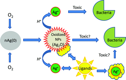
 The released Ag+ is toxic to bacteria due to various mechanisms that include binding to thiol groups in proteins and disrupting their function, compromising membrane permeability leading to cell lysis and death,(17, 18) and oxidative stress due to generation of reactive oxygen species (ROS).(9, 19, 20) However, discerning the contribution of Ag+ vs the AgNPs themselves is challenging due to their common co-occurrence during the exposure period, because most antibacterial assays are conducted under aerobic conditions that promote continuous Ag+ release (Figure 1). Some studies suggest that dissolved Ag+ accounts for most if not all of AgNPs’ toxicity, and AgNPs serve mostly as a source of Ag+.(6, 21) These studies used ligands such as cysteine(21) to neutralize Ag+ and isolate the effect of AgNPs, but this approach may confound the reactivity of the AgNPs themselves due to their potential association with the added ligand. Other studies showed that both AgNPs and Ag+ contribute to the antibacterial activity(7) and toxicity to eukaryotes,(22-26) although their apparent relative importance varies considerably. For example, the toxicity of AgNPs to bacteria (E. coli, B. subtilis, and S. oneidensis)(26) and to the ryegrass Lolium multiflorum(23) can be higher than that exerted by an equivalent Ag+ concentration, although the mechanism responsible for higher toxicity was not discerned.
Figure 1. Role of oxygen and common ligands on the antibacterial activity of AgNPs.
Materials and Methods
Results and Discussion

Figure 2. TEM images of AgNPs stored under (a) anaerobic conditions and (b) aerobic conditions after 10-day exposure to air.

Figure 3. Toxicity of (a) Ag+ and (b) AgNPs under aerobic vs anaerobic conditions. The AgNPs were prepared under anaerobic conditions and tested inside the chamber or outside after exposure to air for 0.5-h (releasing up to 76 μg/L Ag+) or 10-day (releasing up to 181 μg/L Ag+).

Figure 4. Chloride (at equivalent concentrations to the highest silver dose tested) significantly mitigated the toxicity of Ag+ (a), but not AgNPs (b). Exposure to Ag+ was under aerobic conditions with 2.94 μmol/L chloride, whereas exposure to AgNPs was under anaerobic conditions with 57.0 μmol/L chloride.

Figure 5. Equivalent ligand concentrations mitigated the toxicity of Ag+ (2.94 μmol/L) by promoting silver precipitation (with sulfide: 1.47 μmol/L or cysteine: 2.94 μmol/L) or complexation (with chloride: 2.94 μmol/L or phosphate: 0.98 μmol/L) under aerobic conditions.
Acknowledgment
This research was sponsored by a Joint U.S.–U.K. Research Program (U.S.-EPA and U.K.-NERC-ESPRC) (EPA -G2008-STAR-R1). We thank Gautam Kini (Rice University) for his assistance with the particle size and zeta-potential measurements.
Supporting Information
Details on the determination of the minimum lethal concentration (MLC), adherence of AgNPs to polypropylene tubes, and a comparison of Ag+ and ligands concentration with the corresponding solubility product equilibrium constant (Ksp). This material is available free of charge via the Internet at http://pubs.acs.org.
References
This article references 40 other publications.
- 1.Shahverdi
, A. R.; Fakhimi, A.; Shahverdi, H. R.; Minaian, S.Synthesis and effect of silver nanoparticles on the antibacterial activity of different antibiotics against Staphylococcus aureus and Escherichia coli Nanomed.-Nanotechnol. 2007, 3 ( 2) 168– 171[CrossRef], [PubMed], [CAS]1.https://chemport.cas.org/services/resolver?origin=ACS&resolution=options&coi=1%3ACAS%3A528%3ADC%252BD2sXnsVOrurw%253D&md5=8553ebb8071d2379d6b23cf3392df0f9Shahverdi, Ahmad R.; Fakhimi, Ali; Shahverdi, Hamid R.; Minaian, SaraNanomedicine (New York, NY, United States) (2007),Silver nanoparticles (Ag-NPs) have been known to have inhibitory and bactericidal effects. Resistance to antimicrobial agents by pathogenic bacteria has emerged in recent years and is a major health problem. The combination effects of Ag-NPs with the antibacterial activity of antibiotics have not been studied. Here, the authors report on the synthesis of metallic nanoparticles of silver using a redn. of aq. Ag+ ion with the culture supernatants of Klebsiella pneumoniae. These nanoparticles are evaluated for their part in increasing the antimicrobial activities of various antibiotics against Staphylococcus aureus and Escherichia coli. The antibacterial activities of penicillin G, amoxicillin, erythromycin, clindamycin, and vancomycin were increased in the presence of Ag-NPs against both test strains. The highest enhancing effects were obsd. for vancomycin, amoxicillin, and penicillin G against S. aureus. - 2.Shrivastava
, S.; Bera, T.; Roy, A.; Singh, G.; Ramachandrarao, P.; Dash, D.Characterization of enhanced antibacterial effects of novel silver nanoparticles Nanotechnology 2007, 18, ( 22), 225103.[CrossRef], [CAS]2.https://chemport.cas.org/services/resolver?origin=ACS&resolution=options&coi=1%3ACAS%3A528%3ADC%252BD2sXosFSgsbc%253D&md5=d483ba5f141691f9b621f3c8d730e107Shrivastava, Siddhartha; Bera, Tanmay; Roy, Arnab; Singh, Gajendra; Ramachandrarao, P.; Dash, DebabrataNanotechnology (2007),In the present study, we report the prepn. of silver nanoparticles in the range of 10-15 nm with increased stability and enhanced anti-bacterial potency. The morphol. of the nanoparticles was characterized by transmission electron microscopy. The antibacterial effect of silver nanoparticles used in this study was found to be far more potent than that described in the earlier reports. This effect was dose dependent and was more pronounced against gram-neg. bacteria than gram-pos. organisms. Although bacterial cell lysis could be one of the reasons for the obsd. antibacterial property, nanoparticles also modulated the phosphotyrosine profile of putative bacterial peptides, which could thus affect bacterial signal transduction and inhibit the growth of the organisms. - 3.Yoon
, K. Y.; Byeon, J. H.; Park, J. H.; Hwang, J.Susceptibility constants of Escherichia coli and Bacillus subtilis to silver and copper nanoparticles Sci. Total Environ. 2007, 373 ( 2–3) 572– 575[CrossRef], [PubMed], [CAS]3.https://chemport.cas.org/services/resolver?origin=ACS&resolution=options&coi=1%3ACAS%3A528%3ADC%252BD2sXhtlamsLg%253D&md5=8c9cc32b0d2d1a706aff88220da29cf5Yoon, Ki-Young; Byeon, Jeong Hoon; Park, Jae-Hong; Hwang, JunghoScience of the Total Environment (2007),Nanoparticle susceptibility consts. were defined and used to evaluate the antimicrobial characteristics of silver and copper nanoparticles against Escherichia coli and Bacillus subtilis. Reaction of copper nanoparticles of 100 nm with B. subtilis showed the highest susceptibility (Z = 0.0734 mL/μg) whereas the reaction of silver nanoparticles of 40 nm with E. coli showed the lowest one (Z = 0.0236 mL/μg). - 4.Sondi
, I.; Salopek-Sondi, B.Silver nanoparticles as antimicrobial agent: A case study on E-coli as a model for Gram-negative bacteria J. Colloid Interface Sci. 2004, 275 ( 1) 177– 182[CrossRef], [PubMed], [CAS]4.https://chemport.cas.org/services/resolver?origin=ACS&resolution=options&coi=1%3ACAS%3A528%3ADC%252BD2cXktFKktL4%253D&md5=14e07ed929b658a10ddf2bef46114d65Sondi, Ivan; Salopek-Sondi, BrankaJournal of Colloid and Interface Science (2004),The antimicrobial activity of silver nanoparticles against E. coli was investigated as a model for Gram-neg. bacteria. Bacteriol. tests were performed in Luria-Bertani (LB) medium on solid agar plates and in liq. systems supplemented with different concns. of nanosized silver particles. These particles were shown to be an effective bactericide. Scanning and transmission electron microscopy (SEM and TEM) were used to study the biocidal action of this nanoscale material. The results confirmed that the treated E. coli cells were damaged, showing formation of "pits" in the cell wall of the bacteria, while the silver nanoparticles were found to accumulate in the bacterial membrane. A membrane with such a morphol. exhibits a significant increase in permeability, resulting in death of the cell. These nontoxic nanomaterials, which can be prepd. in a simple and cost-effective manner, may be suitable for the formulation of new types of bactericidal materials. - 5.Morones
, J. R.; Elechiguerra, J. L.; Camacho, A.; Holt, K.; Kouri, J. B.; Ramirez, J. T.; Yacaman, M. J.The bactericidal effect of silver nanoparticles Nanotechnology 2005, 16 ( 10) 2346– 2353[CrossRef], [PubMed], [CAS]5.https://chemport.cas.org/services/resolver?origin=ACS&resolution=options&coi=1%3ACAS%3A528%3ADC%252BD2MXht1CiurjJ&md5=116dc1f862d697225bb977a72316963fMorones, Jose Ruben; Elechiguerra, Jose Luis; Camacho, Alejandra; Holt, Katherine; Kouri, Juan B.; Ramirez, Jose Tapia; Yacaman, Miguel JoseNanotechnology (2005),Nanotechnol. is expected to open new avenues to fight and prevent disease using at. scale tailoring of materials. Among the most promising nanomaterials with antibacterial properties are metallic nanoparticles, which exhibit increased chem. activity due to their large surface to vol. ratios and crystallog. surface structure. The study of bactericidal nanomaterials is particularly timely considering the recent increase of new resistant strains of bacteria to the most potent antibiotics. This has promoted research in the well known activity of silver ions and silver-based compds., including silver nanoparticles. The present work studies the effect of silver nanoparticles in the range of 1-100 nm on Gram-neg. bacteria using high angle annular dark field (HAADF) scanning TEM (STEM). Our results indicate that the bactericidal properties of the nanoparticles are size dependent, since the only nanoparticles that present a direct interaction with the bacteria preferentially have a diam. of ∼1-10 nm. - 6.Lok
, C. N.; Ho, C. M.; Chen, R.; He, Q. Y.; Yu, W. Y.; Sun, H.; Tam, P. K. H.; Chiu, J. F.; Che, C. M.Silver nanoparticles: Partial oxidation and antibacterial activities J. Biol. Inorg. Chem. 2007, 12 ( 4) 527– 534[CrossRef], [PubMed], [CAS]6.https://chemport.cas.org/services/resolver?origin=ACS&resolution=options&coi=1%3ACAS%3A528%3ADC%252BD2sXkvVaitLc%253D&md5=226bfec3e8793a41ca274471b985bceaLok, Chun-Nam; Ho, Chi-Ming; Chen, Rong; He, Qing-Yu; Yu, Wing-Yiu; Sun, Hongzhe; Tam, Paul Kwong-Hang; Chiu, Jen-Fu; Che, Chi-MingJBIC, Journal of Biological Inorganic Chemistry (2007),The phys. and chem. properties of silver nanoparticles that are responsible for their antimicrobial activities have been studied with spherical silver nanoparticles (av. diam. approx. 9 nm) synthesized by the borohydride redn. of Ag+ ions, in relation to their sensitivity to oxidn., activities towards silver-resistant bacteria, size-dependent activities, and dispersal in electrolytic solns. Partially (surface) oxidized silver nanoparticles have antibacterial activities, but zero-valent nanoparticles do not. The levels of chemisorbed Ag+ that form on the particle's surface, as revealed by changes in the surface plasmon resonance absorption during oxidn. and redn., correlate well with the obsd. antibacterial activities. Silver nanoparticles, like Ag+ in the form of AgNO3 soln., are tolerated by the bacteria strains resistant to Ag+. The antibacterial activities of silver nanoparticles are related to their size, with the smaller particles having higher activities on the basis of equiv. silver mass content. The silver nanoparticles aggregate in media with a high electrolyte content, resulting in a loss of antibacterial activities. However, complexation with albumin can stabilize the silver nanoparticles against aggregation, leading to a retention of the antibacterial activities. Taken together, the results show that the antibacterial activities of silver nanoparticles are dependent on chemisorbed Ag+, which is readily formed owing to extreme sensitivity to oxygen. The antibacterial activities of silver nanoparticles are dependent on optimally displayed oxidized surfaces, which are present in well-dispersed suspensions. - 7.Fabrega
, J.; Fawcett, S. R.; Renshaw, J. C.; Lead, J. R.Silver nanoparticle impact on bacterial growth: effect of pH, concentration, and organic matter Environ. Sci. Technol. 2009, 43 ( 19) 7285– 7290[ACS Full Text ], [CAS]7.https://chemport.cas.org/services/resolver?origin=ACS&resolution=options&coi=1%3ACAS%3A528%3ADC%252BD1MXmvFCksrw%253D&md5=7546a2fb7aabc9e90bac890e82c7a070Fabrega, Julia; Fawcett, Shona R.; Renshaw, Joanna C.; Lead, Jamie R.Environmental Science & Technology (2009),Ag nanoparticles (NPs) are widely used as antibacterial agents. This antibacterial property carries with it a potential environmental risk once these NPs are discharged into the environment. This study examd. the impact on Pseudomonas fluorescens over a 24 h exposure of well characterized Ag NPs at pH 6-9, in the presence and absence of Suwannee River humic acids (SRHA). Ag NPs were characterized by size, aggregation, morphol., dissoln., and surface properties under all conditions. Soly. was low (<2%) for all Ag NP concns. (2-2000 ppb) and under all conditions was <40 ppb (0.38μM). SRHA caused a partial disaggregation of Ag NP aggregates by nanoscale film formation, with individual NPs stabilized by charge and entropically driven steric effects. Dissolved Ag reduced bacterial growth entirely at 2000 ppb (19μM) under all conditions and adversely affected growth at 200 ppb (1.9μM) under some conditions, indicating some toxicity. The Ag NPs showed similar toxicity at 2000 ppb (19μM) in the absence of SRHA and at pH 9 only i.e. SRHA mitigated bactericidal action. Soly. and interactions with SRHA indicate that there was a specific nanoparticle effect, which could not be explained by the effect of dissolved Ag.
 - 8.Choi
, O.; Deng, K. K.; Kim, N. J.; Ross, L.; Surampalli, R. Y.; Hu, Z. Q.The inhibitory effects of silver nanoparticles, silver ions, and silver chloride colloids on microbial growth Water Res. 2008, 42 ( 12) 3066– 3074[CrossRef], [PubMed], [CAS]8.https://chemport.cas.org/services/resolver?origin=ACS&resolution=options&coi=1%3ACAS%3A528%3ADC%252BD1cXntFGntr8%253D&md5=1148bff38f0c4c9bcf98d55f10c00982Choi, Okkyoung; Deng, Kathy Kanjun; Kim, Nam-Jung; Ross, Louis; Surampalli, Rao Y.; Hu, ZhiqiangWater Research (2008),Emerging nanomaterials are of great concern to wastewater treatment utilities and the environment. The inhibitory effects of silver nanoparticles (Ag NPs) and other important Ag species on microbial growth were evaluated using extant respirometry and an automatic microtiter fluorescence assay. Using autotrophic nitrifying organisms from a well-controlled continuously operated bioreactor, Ag NPs (av. size=14±6 nm), Ag+ ions (AgNO3), and AgCl colloids (av. size = 0.25 μm), all at 1 mg/L Ag, inhibited respiration by 86 ± 3%, 42 ± 7%, and 46 ± 4%, resp. Based on a prolonged microtiter assay, at about 0.5 mg/L Ag, the inhibitions on the growth of Escherichia coli PHL628-gfp by Ag NPs, Ag+ ions, and AgCl colloids were 55 ± 8%, 100%, and 66 ± 6%, resp. Cell membrane integrity was not compromised under the treatment of test Ag species by using a LIVE/DEAD Baclight bacterial viability assay. However, electron micrographs demonstrated that Ag NPs attached to the microbial cells, probably causing cell wall pitting. The results suggest that nitrifying bacteria are esp. susceptible to inhibition by Ag NPs, and the accumulation of Ag NPs could have detrimental effects on the microorganisms in wastewater treatment. - 9.Choi
, O.; Hu, Z. Q.Size dependent and reactive oxygen species related nanosilver toxicity to nitrifying bacteria Environ. Sci. Technol. 2008, 42 ( 12) 4583– 4588[ACS Full Text], [CAS]9.https://chemport.cas.org/services/resolver?origin=ACS&resolution=options&coi=1%3ACAS%3A528%3ADC%252BD1cXlslOjsLk%253D&md5=2fb4b44c8e986176eed13dbc6c72303aChoi, Okkyoung; Hu, ZhiqiangEnvironmental Science & Technology (2008),The intrinsic slow growth of nitrifying bacteria and their high sensitivity to environmental perturbations often result in cell growth inhibition by toxins. Nanoparticles are of great concern to the environment because of their small size and high catalytic properties. This work sought to det. size-dependent inhibition by Ag nanoparticles and evaluate the relationship between the inhibition and reactive oxygen species (ROS). Nanoparticles with an av. size range of 9-21 nm were synthesized by varying the molar ratios of BH4- /Ag+in the soln. The resulting ROS generation was quantified in the presence and absence of the bacteria while the degree of inhibition was inferred from specific oxygen uptake rate measurements, detd. by extant respirometry. By examg. the correlation between nanoparticle size distribution, photocatalytic ROS generation, intracellular ROS accumulation, and nitrification inhibition, we obsd. that inhibition to nitrifying organisms correlated with the fraction of Ag nanoparticles less than 5 nm in the suspension. It appeared that these size nanoparticles could be more toxic to bacteria than any other fractions of nanoparticles or their counterpart bulk species. Furthermore, inhibition by Ag nanoparticles as well as other forms of silver (AgCl colloid and Ag+ ion) correlated well with the intracellular ROS concns., but not with the photocatalytic ROS fractions. The ROS correlations were different for the different forms of silver, indicating that factors other than ROS are also important in detg. nanosilver toxicity. - 10.Pal
, S.; Tak, Y. K.; Song, J. M.Does the antibacterial activity of silver nanoparticles depend on the shape of the nanoparticle? A study of the gram-negative bacterium Escherichia coli Appl. Environ. Microb. 2007, 73 ( 6) 1712– 1720[CrossRef], [PubMed], [CAS]10.https://chemport.cas.org/services/resolver?origin=ACS&resolution=options&coi=1%3ACAS%3A528%3ADC%252BD2sXjs1ahtrk%253D&md5=a7b04e269cac025b87c4151323d9dd41Pal, Sukdeb; Tak, Yu Kyung; Song, Joon MyongApplied and Environmental Microbiology (2007),The authors investigated the antibacterial properties of differently shaped silver nanoparticles against the gram-neg. bacterium Escherichia coli, both in liq. systems and on agar plates. Energy-filtering TEM images revealed considerable changes in the cell membranes upon treatment, resulting in cell death. Truncated triangular silver nanoplates with a {111} lattice plane as the basal plane displayed the strongest biocidal action, compared with spherical and rod-shaped nanoparticles and with Ag+ (in the form of AgNO3). It is proposed that nanoscale size and the presence of a {111} plane combine to promote this biocidal property. To the authors' knowledge, this is the first comparative study on the bactericidal properties of silver nanoparticles of different shapes, and the authors' results demonstrate that silver nanoparticles undergo a shape-dependent interaction with the gram-neg. organism E. coli. - 11.Kim
, J. S.; Kuk, E.; Yu, K. N.; Kim, J. H.; Park, S. J.; Lee, H. J.; Kim, S. H.; Park, Y. K.; Park, Y. H.; Hwang, C. Y.; Kim, Y. K.; Lee, Y. S.; Jeong, D. H.; Cho, M. H.Antimicrobial effects of silver nanoparticles Nanomed.-Nanotechnol. 2007, 3 ( 1) 95– 101[CrossRef], [PubMed], [CAS]11.https://chemport.cas.org/services/resolver?origin=ACS&resolution=options&coi=1%3ACAS%3A528%3ADC%252BD2sXktFCms7s%253D&md5=33a96c57166c125282f194053c49a637Kim, Jun Sung; Kuk, Eunye; Yu, Kyeong Nam; Kim, Jong-Ho; Park, Sung Jin; Lee, Hu Jang; Kim, So Hyun; Park, Young Kyung; Park, Yong Ho; Hwang, Cheol-Yong; Kim, Yong-Kwon; Lee, Yoon-Sik; Jeong, Dae Hong; Cho, Myung-HaingNanomedicine (New York, NY, United States) (2007),The antimicrobial effects of Ag ion or salts are well known, but the effects of Ag nanoparticles on microorganisms and antimicrobial mechanism have not been revealed clearly. Stable Ag nanoparticles were prepd. and their shape and size distribution characterized by particle characterizer and transmission electron microscopic study. The antimicrobial activity of Ag nanoparticles was investigated against yeast, Escherichia coli, and Staphylococcus aureus. In these tests, Muller Hinton agar plates were used and Ag nanoparticles of various concns. were supplemented in liq. systems. Yeast and E. coli were inhibited at the low concn. of Ag nanoparticles, whereas the growth-inhibitory effects on S. aureus were mild. The free-radical generation effect of Ag nanoparticles on microbial growth inhibition was investigated by ESR spectroscopy. These results suggest that Ag nanoparticles can be used as effective growth inhibitors in various microorganisms, making them applicable to diverse medical devices and antimicrobial control systems. - 12.El Badawy
, A. M.; Silva, R. G.; Morris, B.; Scheckel, K. G.; Suidan, M. T.; Tolaymat, T. M.Surface charge-dependent toxicity of silver nanoparticles Environ. Sci. Technol. 2011, 45 ( 1) 283– 7[ACS Full Text], [CAS]12.https://chemport.cas.org/services/resolver?origin=ACS&resolution=options&coi=1%3ACAS%3A528%3ADC%252BC3cXhsFWqsbzL&md5=f975f42464de7f50f8a9f1c16d8ee6b2El Badawy, Amro M.; Silva, Rendahandi G.; Morris, Brian; Scheckel, Kirk G.; Suidan, Makram T.; Tolaymat, Thabet M.Environmental Science & Technology (2011),As a result of the extensive no. of applications of silver nanoparticles (AgNPs), their potential impacts, once released into the environment, are of concern. The toxicity of AgNPs was reported to be dependent on various factors such as particle size, shape and capping agent. Although these factors may play a role in AgNPs toxicity, the results presented herein suggest that surface charge is one of the most important factors that govern the toxicity of AgNPs. In the current study, the toxicity of four AgNPs representing various surface charging scenarios ranging from highly neg. to highly pos. was investigated. These AgNPs were uncoated H2-AgNPs, citrate coated AgNPs (Citrate-AgNPs), polyvinylpyrrolidone coated AgNPs (PVP-AgNPs), and branched polyethyleneimine coated AgNPs (BPEI-AgNPs). The authors' results clearly demonstrate that the AgNPs exhibited surface charge-dependent toxicity on the bacillus species investigated. Furthermore, ultrafiltration membranes were utilized to purify the AgNPs suspensions from residual impurities prior to the introduction to the microbes. This step was crucial in detg. the true AgNPs toxicity and is either missing or not explicitly mentioned in most of the reported toxicity studies. - 13.Rejeski
, D.; Kulken, T.; Pollschuk, P.; Pauwels, E.The project on emerging nanotechnologies. http://www.nanotechproject.org/inventories/consumer/analysis_draft/, 2011.There is no corresponding record for this reference. - 14.Munn
, C. Marine Microbiology; Oxford: BIOS Scientific, 2003.There is no corresponding record for this reference. - 15.Petrucci
, R. H.; Harwood, W. S.; Herring, G. E.; Madura, J. General Chemistry: Principles and Modern Applications, 9th ed.; Prentice Hall: New York, 1997.There is no corresponding record for this reference. - 16.Liu
, J. Y.; Hurt, R. H.Ion release kinetics and particle persistence in aqueous nano-silver colloids Environ. Sci. Technol. 2010, 44 ( 6) 2169– 2175[ACS Full Text], [CAS]16.https://chemport.cas.org/services/resolver?origin=ACS&resolution=options&coi=1%3ACAS%3A528%3ADC%252BC3cXit1Wqsrc%253D&md5=a043ce25387adb8768567c2b35dd76a4Liu, Jingyu; Hurt, Robert H.Environmental Science & Technology (2010),Many important aspects of nanosilver (nAg) are influenced by the ionic activity assocd. with the particle suspension, including antibacterial potency, eukaryotic toxicity, environmental release, and particle persistence. This study synthesizes pure, ion-free, citrate-stabilized nAg colloids as model systems, and measures their time-dependent release of dissolved Ag using centrifugal ultrafiltration and at. absorption spectroscopy. Ion release is shown to be a cooperative oxidn. process requiring both dissolved dioxygen and protons. It produces peroxide intermediates, and proceeds to complete reactive dissoln. under some conditions. Ion release rates increase with temp. (0-37°), and decrease with increasing pH or addn. of humic or fulvic acids. Sea salts have only a minor effect on dissolved Ag release. Ag nanoparticle surfaces can adsorb Ag+, so even simple colloids contain 3 forms of Ag: Ag0 solids, free Ag+ or its complexes, and surface-adsorbed Ag+. Both thermodn. anal. and kinetic measurements indicate that Ag0 nanoparticles will not be persistent in realistic environmental compartments contg. dissolved O. An empirical kinetic law is proposed that reproduces the obsd. effects of dissoln. time, pH, humic/fulvic acid content, and temp. obsd. here in the low range of nAg concn. most relevant for the environment. - 17.Ratte
, H. T.Bioaccumulation and toxicity of silver compounds: A review Environ. Toxicol. Chem. 1999, 18 ( 1) 89– 108[CrossRef], [CAS]17.https://chemport.cas.org/services/resolver?origin=ACS&resolution=options&coi=1%3ACAS%3A528%3ADyaK1MXkt1Or&md5=4f96982bc1c71fffb5ceb4fc76bf004eRatte, Hans ToniEnvironmental Toxicology and Chemistry (1999),A review with 123 refs. of the literature revealed that bioaccumulation of silver in soil is rather low, even if the soil is amended with silver-contg. sewage sludge. Plants grown on tailings of silver mines were found to have silver primarily in the root systems. In marine and freshwater systems, the highest reported bioconcn. factors (BCFs) were obsd. in algae (>105), probably because of adsorption of the dissolved silver (<0.45 p.m fraction) to the cell surface. In herbivorous organisms (e.g., zooplankton and bivalves), the BCF was lower by about two orders of magnitude. Low amts. of silver were assimilated from food with no substantial biomagnification. In carnivores (e.g., fish), the BCF was also lower by one order of magnitude with no indication of biomagnification. Toxicity of silver occurs mainly in the aq. phase and depends on the concn. of active, free Ag+ ions. Accordingly, many processes and water characteristics reduce silver toxicity by stopping the formation of free Ag-, binding Ag+, or preventing binding of Ag+ to the reactive surfaces of organisms. The soly. of a silver compd., and the presence of complexing agents (e.g., thiosulfate or chloride), dissolved org. carbon, and competing ions are important. In soil. sewage sludge, and sediments, in which silver sulfide predominates, the toxicity of silver, even at high total concns., is very low. The highly sol. silver thiosulfate complex has low toxicity, which can be attributed to the silver complexed by thiosulfate. Silver nitrate is one of the most toxic silver compds. The toxic potential of silver chloride complexes in seawater is and will be an important issue for investigation. Aquatic chronic tests, long-term tests, and tests including sensitive life stages show lower toxicity thresholds (∼1 μg Ag+/L). The organisms viewed as most sensitive to silver are small aquatic invertebrates, particularly embryonic and larval stages. - 18.Wang
, J. M.; Huang, C. P.; Pirestani, D.Interactions of silver with wastewater constituents Water Res. 2003, 37 ( 18) 4444– 4452[CrossRef], [PubMed], [CAS]18.https://chemport.cas.org/services/resolver?origin=ACS&resolution=options&coi=1%3ACAS%3A528%3ADC%252BD3sXntl2jur0%253D&md5=ce994664ead3ccb9a31fbd03988d3bd0Wang, Jianmin; Huang, C. P.; Pirestani, D.Water Research (2003),The interactions of Ag ion with wastewater constituents including chloride, sludge particulates, and dissolved org. matter (DOM) were investigated using fresh and processed sludge samples from various municipal wastewater treatment plants. Fresh sludge samples were used to examine the Ag removal characteristics in the presence of chloride. Processed sludge samples that were subjected to a deionized-water washing procedure to remove the chloride ion were used to examine the interactions among Ag(I), sludge solids, and DOM. Results showed that, under field conditions, most of the Ag can be removed through pptn. with chloride and adsorption by sludge particulates. Results obtained using washed sludge samples showed that Ag(I) adsorption by sludge particulates increases with the increase of pH in the acidic pH range, but decreases with the increase of pH in the alk. pH range. Since the DOM concn. increases significantly with the increase of pH in the alk. pH region, it can be concluded that the formation of Ag(I)-DOM complexes reduces the Ag(I) adsorption by sludge. Fitting of the Langmuir isotherm to the adsorption data indicated that adsorption consts. have a slight increase with the increase of pH in the acidic pH range, but decrease significantly with the increase of pH in the alk. pH range. The decrease of the Ag(I) adsorption consts. in the alk. pH range can be attributed to the formation of Ag(I)-DOM complexes which weaken the Ag(I) binding strength by sludge surface sites. The fitted isotherms also showed that the Ag(I) adsorption capacity increases with the increase of pH in the entire pH range. - 19.Kim
, S.; Choi, J. E.; Choi, J.; Chung, K. H.; Park, K.; Yi, J.; Ryu, D. Y.Oxidative stress-dependent toxicity of silver nanoparticles in human hepatoma cells Toxicol. in Vitro 2009, 23 ( 6) 1076– 1084[CrossRef], [PubMed], [CAS]19.https://chemport.cas.org/services/resolver?origin=ACS&resolution=options&coi=1%3ACAS%3A528%3ADC%252BD1MXps1Cnurc%253D&md5=73f986dde53813a9475921df2e7bca08Kim, Soohee; Choi, Ji Eun; Choi, Jinhee; Chung, Kyu-Hyuck; Park, Kwangsik; Yi, Jongheop; Ryu, Doug-YoungToxicology in Vitro (2009),Cytotoxicity induced by silver nanoparticles (AgNPs) and the role that oxidative stress plays in this process were demonstrated in human hepatoma cells. Toxicity induced by silver (Ag+) ions was studied in parallel using AgNO3 as the Ag+ ion source. Using cation exchange treatment, we confirmed that the AgNP soln. contained a negligible amt. of free Ag+ ions. Metal-responsive metallothionein 1b (MT1b) mRNA expression was not induced in AgNP-treated cells, while it was induced in AgNO3-treated cells. These results indicate that AgNP-treated cells have limited exposure to Ag+ ions, despite the potential release of Ag+ ions from AgNPs in cell culture. AgNPs agglomerated in the cytoplasm and nuclei of treated cells, and induced intracellular oxidative stress. AgNPs exhibited cytotoxicity with a potency comparable to that of Ag+ ions in in vitro cytotoxicity assays. However, the toxicity of AgNPs was prevented by use of the antioxidant N-acetylcysteine, and AgNP-induced DNA damage was also prevented by N-acetylcysteine. AgNO3 treatment induced oxidative stress-related glutathione peroxidase 1 (GPx1) and catalase expression to a greater extent than AgNP exposure, but treatment with AgNO3 and AgNPs induced comparable superoxide dismutase 1 (SOD1) expression levels. Our findings suggest that AgNP cytotoxicity is primarily the result of oxidative stress and is independent of the toxicity of Ag+ ions. - 20.Park
, H. J.; Kim, J. Y.; Kim, J.; Lee, J. H.; Hahn, J. S.; Gu, M. B.; Yoon, J.Silver-ion-mediated reactive oxygen species generation affecting bactericidal activity Water Res. 2009, 43 ( 4) 1027– 1032[CrossRef], [PubMed], [CAS]20.https://chemport.cas.org/services/resolver?origin=ACS&resolution=options&coi=1%3ACAS%3A528%3ADC%252BD1MXisVGjsbs%253D&md5=5891f2edb542284e7f995643f34e2168Park, Hee-Jin; Kim, Jee Yeon; Kim, Jaeeun; Lee, Joon-Hee; Hahn, Ji-Sook; Gu, Man Bock; Yoon, JeyongWater Research (2009),Silver ions have been widely used as disinfectants that inhibit bacterial growth by inhibiting the essential enzymic functions of the microorganism via interaction with the thiol-group of L-cysteine. However, silver-ion-mediated perturbation of the bacterial respiratory chain has raised the possibility of reactive oxygen species (ROS) generation. We used bacterial reporter strains specifically responding to superoxide radicals and found that silver-ion-mediated ROS-generation affected bactericidal activity. Almost half the log redn. in Escherichia coli and Staphylococcus aureus populations (model strains for gram neg. and pos. bacteria, resp.) caused by silver-ion disinfection was attributed to ROS-mediated bactericidal activity. The major form of ROS generated was the superoxide-radical; H2O2 was not induced. Furthermore, silver ions strongly enhanced paraquat-induced oxidative stress, indicating close correlation and synergism between the conventional and ROS-mediated silver toxicity. Our results suggest that further studies in silver-based disinfection systems should consider the oxygen concn. and ROS reaction. - 21.Navarro
, E.; Piccapietra, F.; Wagner, B.; Marconi, F.; Kaegi, R.; Odzak, N.; Sigg, L.; Behra, R.Toxicity of Silver Nanoparticles to Chlamydomonas reinhardtii Environ. Sci. Technol. 2008, 42 ( 23) 8959– 8964[ACS Full Text], [CAS]21.https://chemport.cas.org/services/resolver?origin=ACS&resolution=options&coi=1%3ACAS%3A528%3ADC%252BD1cXhtFOqt7nO&md5=da32b6e9fb221d6c2943e9388ea374cfNavarro, Enrique; Piccapietra, Flavio; Wagner, Bettina; Marconi, Fabio; Kaegi, Ralf; Odzak, Niksa; Sigg, Laura; Behra, RenataEnvironmental Science & Technology (2008),Silver nanoparticles (AgNP) are likely to enter the aquatic environment because of their multiple uses. The authors have examd. the short-term toxicity of AgNP and ionic silver (Ag+) to photosynthesis in C. reinhardtii using fluorometry. AgNP ranged in size from 10 to 200 nm with most particles around 25 nm. As detd. by DGT (diffusive gradients in thin films), by ion-selective electrode, and by centrifugal ultrafiltration, ∼1% of the AgNP was present as Ag+ ions. Based on total Ag concn., toxicity was 18 times higher for AgNO3 than for AgNP (in terms of EC50). However, when compared as a function of the Ag+ concn., the toxicity of AgNP appeared to be much higher than that of AgNO3. The ionic Ag+ measured in the AgNP suspensions could not fully explain the obsd. toxicity. Cysteine, a strong Ag+ ligand, abolished the inhibitory effects on photosynthesis of both AgNP and Ag+. Together, the results indicate that the interaction of these particles with algae influences the toxicity of AgNP, which is mediated by Ag+. Particles contributed to the toxicity as a source of Ag+ which is formed in the presence of algae. - 22.Meyer
, J. N.; Lord, C. A.; Yang, X. Y. Y.; Turner, E. A.; Badireddy, A. R.; Marinakos, S. M.; Chilkoti, A.; Wiesner, M. R.; Auffan, M.Intracellular uptake and associated toxicity of silver nanoparticles in Caenorhabditis elegans Aquat. Toxicol. 2010, 100 ( 2) 140– 150[CrossRef], [PubMed], [CAS]22.https://chemport.cas.org/services/resolver?origin=ACS&resolution=options&coi=1%3ACAS%3A528%3ADC%252BC3cXhtFGjs7bI&md5=f43396ceff0affd43f9f247b2624fb6bMeyer, Joel N.; Lord, Christopher A.; Yang, Xinyu Y.; Turner, Elena A.; Badireddy, Appala R.; Marinakos, Stella M.; Chilkoti, Ashutosh; Wiesner, Mark R.; Auffan, MelanieAquatic Toxicology (2010),Silver nanoparticles (AgNPs) are frequently used as antimicrobials. While the mechanism(s) by which AgNPs are toxic are unclear, their increasing use raises the concern that release into the environment could lead to environmental toxicity. We characterized the physicochem. behavior, uptake, toxicity (growth inhibition), and mechanism of toxicity of three AgNPs with different sizes and polyvinylpyrrolidone (PVP) or citrate coatings to the nematode Caenorhabditis elegans. We used wild-type (N2) C. elegans and strains expected to be sensitive to oxidative stress (nth-1, sod-2 and mev-1), genotoxins (xpa-1 and nth-1), and metals (mtl-2). Using traditional and novel anal. methods, we obsd. significant aggregation and extra-organismal dissoln. of silver, organismal uptake and, in one case, transgenerational transfer of AgNPs. We also obsd. growth inhibition by all tested AgNPs at concns. in the low mg/L levels. A metallothionein-deficient (mtl-2) strain was the only mutant tested that exhibited consistently greater AgNP sensitivity than wild-type. Although all tested AgNPs were internalized (passed cell membranes) in C. elegans, at least part of the toxicity obsd. was mediated by ionic silver. Finally, we describe a modified growth assay that permits differentiation between direct growth-inhibitory effects and indirect inhibition mediated by toxicity to the food source. - 23.Yin
, L. Y.; Cheng, Y. W.; Espinasse, B.; Colman, B. P.; Auffan, M.; Wiesner, M.; Rose, J.; Liu, J.; Bernhardt, E. S.More than the ions: the effects of silver nanoparticles on Lolium multiflorum Environ. Sci. Technol. 2011, 45 ( 6) 2360– 2367[ACS Full Text], [CAS]23.https://chemport.cas.org/services/resolver?origin=ACS&resolution=options&coi=1%3ACAS%3A528%3ADC%252BC3MXitlamtLk%253D&md5=cac8a722359eaab83152e01777337942Yin, Liyan; Cheng, Yingwen; Espinasse, Benjamin; Colman, Benjamin P.; Auffan, Melanie; Wiesner, Mark; Rose, Jerome; Liu, Jie; Bernhardt, Emily S.Environmental Science & Technology (2011),Silver nanoparticles (AgNPs) are increasingly used as antimicrobial additives in consumer products and may have adverse impacts on organisms when they inadvertently enter ecosystems. This study investigated the uptake and toxicity of AgNPs to the common grass, L. multiflorum. Root and shoot Ag content increased with increasing AgNP exposures. AgNPs inhibited seedling growth. While exposed to 40 mg L-1 gum arabic-coated AgNPs, seedlings failed to develop root hairs, had highly vacuolated and collapsed cortical cells, and broken epidermis and root cap. In contrast, seedlings exposed to identical concns. of AgNO3 or supernatants of ultracentrifuged AgNP solns. showed no such abnormalities. AgNP toxicity was influenced by total NP surface area with smaller AgNPs (6 nm) more strongly affecting growth than did similar concns. of larger (25 nm) NPs for a given mass. Cysteine (which binds Ag+) mitigated the effects of AgNO3 but did not reduce the toxicity of AgNP treatments. X-ray spectromicroscopy documented silver speciation within exposed roots and suggested that silver is oxidized within plant tissues. Collectively, this study suggests that growth inhibition and cell damage can be directly attributed either to the nanoparticles themselves or to the ability of AgNPs to deliver dissolved Ag to crit. biotic receptors. - 24.Kawata
, K.; Osawa, M.; Okabe, S.In vitro toxicity of silver nanoparticles at noncytotoxic doses to HepG2 human hepatoma cells Environ. Sci. Technol. 2009, 43 ( 15) 6046– 6051[ACS Full Text], [CAS]24.https://chemport.cas.org/services/resolver?origin=ACS&resolution=options&coi=1%3ACAS%3A528%3ADC%252BD1MXot12msrk%253D&md5=868eb20dccdfd3bf819b03352a08e54dKawata, Koji; Osawa, Masato; Okabe, SatoshiEnvironmental Science & Technology (2009),Although it has been reported that silver nanoparticles (Ag-NPs) have strong acute toxic effects to various cultured cells, the toxic effects at noncytotoxic doses are still unknown. We, therefore, evaluated in vitro toxicity of Ag-NPs at noncytotoxic doses in human hepatoma cell line, HepG2, based on cell viability assay, micronucleus test, and DNA microarray anal. We also used polystyrene nanoparticles (PS-NPs) and silver carbonate (Ag2CO3) as test materials to compare the toxic effects with respect to different raw chem. compn. and form of silver. The cell viability assay demonstrated that Ag-NPs accelerated cell proliferation at low doses (<0.5 mg/L), which was supported by the DNA microarray anal. showing significant induction of genes assocd. with cell cycle progression. However, only Ag-NPs exposure exhibited a significant cytotoxicity at higher doses (>1.0 mg/L) and induced abnormal cellular morphol., displaying cellular shrinkage and acquisition of an irregular shape. In addn., only Ag-NPs exposure increased the frequency of micronucleus formation up to 47.9 ± 3.2% of binucleated cells, suggesting that Ag-NPs appear to cause much stronger damages to chromosome than PS-NPs and ionic Ag+. Cysteine, a strong ionic Ag+ ligand, only partially abolished the formation of micronuclei mediated by Ag-NPs and changed the gene expression, indicating that ionic Ag+ derived from Ag-NPs could not fully explain these biol. actions. Based on these discussions, it is concluded that both "nanosized particle of Ag" as well as "ionic Ag+" contribute to the toxic effects of Ag-NPs. - 25.Laban
, G.; Nies, L. F.; Turco, R. F.; Bickham, J. W.; Sepulveda, M. S.The effects of silver nanoparticles on fathead minnow (Pimephales promelas) embryos Ecotoxicology 2010, 19 ( 1) 185– 195[CrossRef], [PubMed], [CAS]25.https://chemport.cas.org/services/resolver?origin=ACS&resolution=options&coi=1%3ACAS%3A528%3ADC%252BC3cXitlKgsQ%253D%253D&md5=25f03912c78d11529df8c39bc781c544Laban, Geoff; Nies, Loring F.; Turco, Ronald F.; Bickham, John W.; Sepulveda, Maria S.Ecotoxicology (2010),Nanoparticles are being used in many com. applications. We describe the toxicity of two com. silver (Ag) nanoparticle (NP) products, NanoAmor and Sigma on Pimephales promelas embryos. Embryos were exposed to varying concns. of either sonicated or stirred NP solns. for 96 h. LC50 values for NanoAmor and Sigma Ag NPs were 9.4 and 10.6 mg/L for stirred and 1.25 and 1.36 mg/L for sonicated NPs, resp. Uptake of Ag NPs into the embryos was obsd. after 24 h using Transmission Electron Microscopy and Ag NPs induced a concn.-dependent increase in larval abnormalities, mostly edema. Dissolved Ag released from Ag NPs was measured using Inductively Coupled-Mass Spectrometry and the effects tested were found to be three times less toxic when compared to Ag nitrate (AgNO3). The percentage of dissolved Ag released was inversely proportional to the concn. of Ag NPs with the lowest (0.625 mg/L) and highest (20 mg/L) concns. tested releasing 3.7 and 0.45% dissolved Ag, resp. and percent release was similar regardless if concns. were stirred or sonicated. Thus increased toxicity after sonication cannot be solely explained by dissolved Ag. We conclude that both dissolved and particulate forms of Ag elicited toxicity to fish embryos. - 26.Suresh
, A. K.; Pelletier, D. A.; Wang, W.; Moon, J. W.; Gu, B. H.; Mortensen, N. P.; Allison, D. P.; Joy, D. C.; Phelps, T. J.; Doktycz, M. J.Silver nanocrystallites: biofabrication using Shewanella oneidensis, and an evaluation of their comparative toxicity on gram-negative and gram-positive bacteria Environ. Sci. Technol. 2010, 44 ( 13) 5210– 5215[ACS Full Text], [CAS]26.https://chemport.cas.org/services/resolver?origin=ACS&resolution=options&coi=1%3ACAS%3A528%3ADC%252BC3cXmslemtLk%253D&md5=332d316b2a9bb4b53a10940754ccadd3Suresh, Anil K.; Pelletier, Dale A.; Wang, Wei; Moon, Ji-Won; Gu, Baohua; Mortensen, Ninell P.; Allison, David P.; Joy, David C.; Phelps, Tommy J.; Doktycz, Mitchel J.Environmental Science & Technology (2010),Microorganisms have long been known to develop resistance to metal ions either by sequestering metals inside the cell or by effluxing them into the extracellular media. Here, the authors report the biosynthesis of extracellular silver-based single nanocrystallites of well defined compn. and homogeneous morphol. utilizing the γ-proteobacterium Shewanella oneidensis MR-1 upon incubation with aq. silver nitrate soln. Further characterization of these particles revealed that the crystals consist of small, reasonably monodispersed spheres in the 2-11 nm size range (av. of 4 ± 1.5 nm). The bactericidal effect of these nanoparticles (biogenic-Ag) is compared to chem. synthesized silver nanoparticles (colloidal-Ag and oleate capped silver nanoparticles, oleate-Ag) and assessed using Gram-neg. (E. coli and S. oneidensis) and Gram-pos. (B. subtilis) bacteria. Relative toxicity was based on the diam. of inhibition zone in disk diffusion tests, min. inhibitory concns., live/dead assays, and at. force microscopy. From a toxicity perspective, strain-dependent inhibition depended on the synthesis procedure and the surface coat. Biogenic-Ag was found to be of higher toxicity compared to colloidal-Ag for all three strains tested, whereas E. coli and S. oneidensis were found to be more resistant to either of these nanoparticles than B. subtilis. In contrast, oleate-Ag was not toxic to any of the bacteria. These findings have implications for the potential uses of Ag nanomaterials and for their fate in biol. and environmental systems. - 27.Sotiriou
, G. A.; Pratsinis, S. E.Antibacterial activity of nanosilver ions and particles Environ. Sci. Technol. 2010, 44 ( 14) 5649– 5654[ACS Full Text], [CAS]27.https://chemport.cas.org/services/resolver?origin=ACS&resolution=options&coi=1%3ACAS%3A528%3ADC%252BC3cXotVers7o%253D&md5=6f88efa1948690ac51f5ed3698e5850cSotiriou, Georgios A.; Pratsinis, Sotiris E.Environmental Science & Technology (2010),The antibacterial activity of nanosilver against Gram neg. E. coli is studied by immobilizing nanosilver on nanostructured silica particles and closely controlling Ag content and size. These Ag/SiO2 nanoparticles were characterized by S/TEM, EDX spectroscopy, x-ray diffraction the exposed Ag surface area was measured qual. by O chemisorption. The fraction of dissolved nanosilver was detd. by measuring the released (leached) Ag+ concn. in aq. suspensions of such Ag/SiO2 particles. The antibacterial effect of Ag+ was distinguished from that of nanosilver particles by monitoring the growth of E. coli populations in the presence and absence of Ag/SiO2 particles. The antibacterial activity of nanosilver was dominated by Ag+ when fine Ag nanoparticles (.ltorsim.10 nm in av. diam.) were used that release high concns. of Ag+. In contrast, when relatively larger Ag nanoparticles were used, the concn. of the released Ag+ was lower. Then the antibacterial activity of the released Ag+ and nanosilver particles was comparable. - 28.Panacek
, A.; Kvitek, L.; Prucek, R.; Kolar, M.; Vecerova, R.; Pizurova, N.; Sharma, V. K.; Nevecna, T.; Zboril, R.Silver colloid nanoparticles: synthesis, characterization, and their antibacterial activity J. Phys. Chem. B 2006, 110 ( 33) 16248– 16253[ACS Full Text], [CAS]28.https://chemport.cas.org/services/resolver?origin=ACS&resolution=options&coi=1%3ACAS%3A528%3ADC%252BD28XnsFemt7s%253D&md5=d780f35f8a83f7a856ad4e084f9719b3Panacek, Ales; Kvitek, Libor; Prucek, Robert; Kolar, Milan; Vecerova, Renata; Pizurova, Nadezda; Sharma, Virender K.; Nevecna, Tat'jana; Zboril, RadekJournal of Physical Chemistry B (2006),A one-step simple synthesis of silver colloid nanoparticles with controllable sizes is presented. In this synthesis, redn. of [Ag(NH3)2]+ complex cation by four saccharides was performed. Four saccharides were used: two monosaccharides (glucose and galactose) and two disaccharides (maltose and lactose). The syntheses performed at various ammonia concns. (0.005-0.20 mol L-1) and pH conditions (11.5-13.0) produced a wide range of particle sizes (25-450 nm) with narrow size distributions, esp. at the lowest ammonia concns. The av. size, size distribution, morphol., and structure of particles were detd. by dynamic light scattering (DLS), transmission electron microscopy (TEM), and UV/Visible absorption spectrophotometry. The influence of the saccharide structure (monosaccharides vs. disaccharides) on the size of silver particles is briefly discussed. The redn. of [Ag(NH3)2]+ by maltose produced silver particles with a narrow size distribution with an av. size of 25 nm, which showed high antimicrobial and bactericidal activity against Gram-pos. and Gram-neg. bacteria, including highly multiresistant strains such as methicillin-resistant Staphylococcus aureus. Antibacterial activity of silver nanoparticles was found to be dependent on the size of silver particles. A very low concn. of silver (as low as 1.69 μg/mL Ag) gave antibacterial performance. - 29.Carlson
, C.; Hussain, S. M.; Schrand, A. M.; Braydich-Stolle, L. K.; Hess, K. L.; Jones, R. L.; Schlager, J. J.Unique cellular interaction of silver nanoparticles: size-dependent generation of reactive oxygen species J. Phys. Chem. B 2008, 112 ( 43) 13608– 13619[ACS Full Text], [CAS]29.https://chemport.cas.org/services/resolver?origin=ACS&resolution=options&coi=1%3ACAS%3A528%3ADC%252BD1cXhtF2gs7jP&md5=85ca010191f0a37a7c65f4886a699ff6Carlson, C.; Hussain, S. M.; Schrand, A. M.; Braydich-Stolle, L. K.; Hess, K. L.; Jones, R. L.; Schlager, J. J.Journal of Physical Chemistry B (2008),The rapid advancement of nanotechnol. has created a vast array of engineered nanomaterials (ENMs) which have unique phys. (size, shape, crystallinity, surface charge) and chem. (surface coating, elemental compn. and soly.) attributes. These physicochem. properties of ENMs can produce chem. conditions to induce a pro-oxidant environment in the cells, causing an imbalanced cellular energy system dependent on redox potential and thereby leading to adverse biol. consequences, ranging from the initiation of inflammatory pathways through to cell death. The present study was designed to evaluate size-dependent cellular interactions of known biol. active silver nanoparticles (NPs, Ag-15nm, Ag-30nm, and Ag-55nm). Alveolar macrophages provide the first defense and were studied for their potential role in initiating oxidative stress. Cell exposure produced morphol. abnormal sizes and adherence characteristics with significant NP uptake at high doses after 24 h. Toxicity evaluations using mitochondrial and cell membrane viability along with reactive oxygen species (ROS) were performed. After 24 h of exposure, viability metrics significantly decreased with increasing dose (10-75 μg/mL) of Ag-15nm and Ag-30nm NPs. A more than 10-fold increase of ROS levels in cells exposed to 50 μg/mL Ag-15nm suggests that the cytotoxicity of Ag-15nm is likely to be mediated through oxidative stress. In addn., activation of the release of traditional inflammatory mediators were examd. by measuring levels of cytokines/chemokines, including tumor necrosis factor (TNF-α), macrophage inhibitory protein (MIP-2), and interleukin-6 (IL-6), released into the culture media. After 24 h of exposure to Ag-15nm nanoparticles, a significant inflammatory response was obsd. by the release of TNF-α, MIP-2, and IL-1β. However, there was no detectable level of IL-6 upon exposure to silver nanoparticles. In summary, a size-dependent toxicity was produced by silver nanoparticles, and one predominant mechanism of toxicity was found to be largely mediated through oxidative stress. - 30.Choi
, O.; Cleuenger, T. E.; Deng, B. L.; Surampalli, R. Y.; Ross, L.; Hu, Z. Q.Role of sulfide and ligand strength in controlling nanosilver toxicity Water Res. 2009, 43 ( 7) 1879– 1886[CrossRef], [PubMed], [CAS]30.https://chemport.cas.org/services/resolver?origin=ACS&resolution=options&coi=1%3ACAS%3A528%3ADC%252BD1MXktVCmtb0%253D&md5=b809776e2ed47c2fde174c85b5b73dc0Choi, Okkyoung; Clevenger, Thomas E.; Deng, Baolin; Surampalli, Rao Y.; Ross, Louis; Hu, ZhiqiangWater Research (2009),Nanosilver has been used broadly in nanotechnol. enhanced consumer products because of its strong antimicrobial properties. Silver nanoparticles (AgNPs) released from these products will likely enter wastewater collection and treatment systems. This research evaluated the role of sulfide and ligand strength in controlling nanosilver toxicity to nitrifying bacteria that are important in wastewater treatment. The nanosilver toxicity in the absence and presence of ligands (SO42-, S2-, Cl-, PO43-, and EDTA-) commonly present in wastewater was detd. from the oxygen uptake rate measurements. Sulfide appeared to be the only ligand to effectively reduce nanosilver toxicity. By adding a small aliquot of sulfide that was stoichiometrically complexed with AgNPs, the nanosilver toxicity to nitrifying organisms was reduced by up to 80%. SEM coupled with energy dispersive X-ray anal. indicated that AgNPs were highly reactive with sulfide to form new AgxSy complexes or ppts. These complexes were not oxidized after a prolonged period of aeration (18 h). This information is useful for wastewater treatment design and operation to reduce nanosilver toxicity via sulfide complexation. While the biotic ligand model was successful in predicting the toxicity of Ag+ ions, it could not accurately predict the toxicity of AgNPs. Nevertheless, it could be one of the many tools useful in predicting and controlling nanosilver toxicity to wastewater microorganisms. - 31.Zodrow
, K.; Brunet, L.; Mahendra, S.; Li, D.; Zhang, A.; Li, Q. L.; Alvarez, P. J. J.Polysulfone ultrafiltration membranes impregnated with silver nanoparticles show improved biofouling resistance and virus removal Water Res. 2009, 43 ( 3) 715– 723[CrossRef], [PubMed], [CAS]31.https://chemport.cas.org/services/resolver?origin=ACS&resolution=options&coi=1%3ACAS%3A528%3ADC%252BD1MXhsFelsb8%253D&md5=ec610b7c06515992084af9ada3d0259dZodrow, Katherine; Brunet, Lena; Mahendra, Shaily; Li, Dong; Zhang, Anna; Li, Qilin; Alvarez, Pedro J. J.Water Research (2009),Biofouling and virus penetration are two significant obstacles in water treatment membrane filtration. Biofouling reduces membrane permeability, increases energy costs, and decreases the lifetime of membranes. In order to effectively remove viruses, nanofiltration or reverse osmosis (both high energy filtration schemes) must be used. Thus, there is an urgent demand for low pressure membranes with anti-biofouling and antiviral properties. The antibacterial properties of silver are well known, and silver nanoparticles (nAg) are now incorporated into a wide variety of consumer products for microbial control. In this study, nAg incorporated into polysulfone ultrafiltration membranes (nAg-PSf) exhibited antimicrobial properties towards a variety of bacteria, including Escherichia coli K12 and Pseudomonas mendocina KR1, and the MS2 bacteriophage. Nanosilver incorporation also increased membrane hydrophilicity, reducing the potential for other types of membrane fouling. XPS anal. indicated a significant loss of silver from the membrane surface after a relatively short filtration period (0.4 L/cm2) even though ICP anal. of digested membrane material showed that 90% of the added silver remained in the membrane. This silver loss resulted in a significant loss of antibacterial and antiviral activity. Thus, successful fabrication of nAg-impregnated membranes needs to allow for the release of sufficient silver ions for microbial control while preventing a rapid depletion of silver. - 32.Andrews
, J. M.Determination of minimum inhibitory concentrations J. Antimicrob. Chemoth. 2001, 48, 5– 16[CrossRef], [PubMed], [CAS]32.https://chemport.cas.org/services/resolver?origin=ACS&resolution=options&coi=1%3ACAS%3A528%3ADC%252BD3MXlsVegtr8%253D&md5=8eb739d532e5045be7b376d586002d96Andrews, Jennifer M.Journal of Antimicrobial Chemotherapy (2001),A review with 3 refs. Min. inhibitory concns. (MICs) are defined as the lowest concn. of an antimicrobial that will inhibit the visible growth of a microorganism after overnight incubation, and min. bactericidal concns. (MBCs) as the lowest concn. of antimicrobial that will prevent the growth of an organism after subculture on to antibiotic-free media. MICs are used by diagnostic labs. mainly to confirm resistance, but most often as a research tool to det. the in vitro activity of new antimicrobials, and data from such studies have been used to det. MIC breakpoints. MBC detns. are undertaken less frequently and their major use has been reserved for isolates from the blood of patients with endocarditis. Standardized methods for detg. MICs and MBCs are described in this paper. Like all standardized procedures, the method must be adhered to and may not be adapted by the user. The method gives information on the storage of std. antibiotic powder, prepn. of stock antibiotic solns., media, prepn. of inocula, incubation conditions, and reading and interpretation of results. Tables giving expected MIC ranges for control NCTC and ATCC strains are also supplied. - 33.Adams
, L. K.; Lyon, D. Y.; Alvarez, P. J. J.Comparative eco-toxicity of nanoscale TiO2, SiO2, and ZnO water suspensions Water Res. 2006, 40 ( 19) 3527– 3532[CrossRef], [PubMed], [CAS]33.https://chemport.cas.org/services/resolver?origin=ACS&resolution=options&coi=1%3ACAS%3A528%3ADC%252BD28XhtVOgt7fN&md5=fa499d34b8042087f8d2499d11993784Adams, Laura K.; Lyon, Delina Y.; Alvarez, Pedro J. J.Water Research (2006),The potential eco-toxicity of nanosized titanium dioxide (TiO2), silicon dioxide (SiO2), and zinc oxide (ZnO) water suspensions was investigated using Gram-pos. Bacillus subtilis and Gram-neg. Escherichia coli as test organisms. These three photosensitive nanomaterials were harmful to varying degrees, with antibacterial activity increasing with particle concn. Antibacterial activity generally increased from SiO2 to TiO2 to ZnO, and B. subtilis was most susceptible to their effects. Advertized nanoparticle size did not correspond to true particle size. Apparently, aggregation produced similarly sized particles that had similar antibacterial activity at a given concn. The presence of light was a significant factor under most conditions tested, presumably due to its role in promoting generation of reactive oxygen species (ROS). However, bacterial growth inhibition was also obsd. under dark conditions, indicating that undetd. mechanisms addnl. to photocatalytic ROS prodn. were responsible for toxicity. These results highlight the need for caution during the use and disposal of such manufd. nanomaterials to prevent unintended environmental impacts, as well as the importance of further research on the mechanisms and factors that increase toxicity to enhance risk management. - 34.Liu
, J. Y.; Sonshine, D. A.; Shervani, S.; Hurt, R. H.Controlled release of biologically active silver from nanosilver surfaces ACS Nano 2010, 4 ( 11) 6903– 6913[ACS Full Text], [CAS]34.https://chemport.cas.org/services/resolver?origin=ACS&resolution=options&coi=1%3ACAS%3A528%3ADC%252BC3cXhtlaitb3L&md5=3e3b976bfa5f17404e478b06e4d55febLiu, Jingyu; Sonshine, David A.; Shervani, Saira; Hurt, Robert H.ACS Nano (2010),Major pathways in the antibacterial activity and eukaryotic toxicity of nanosilver involve the silver cation and its sol. complexes, which are well established thiol toxicants. Through these pathways, nanosilver behaves in analogy to a drug delivery system, in which the particle contains a concd. inventory of an active species, the ion, which is transported to and released near biol. target sites. Although the importance of silver ion in the biol. response to nanosilver is widely recognized, the drug delivery paradigm has not been well developed for this system, and there is significant potential to improve nanosilver technologies through controlled release formulations. This article applies elements of the drug delivery paradigm to nanosilver dissoln. and presents a systematic study of chem. concepts for controlled release. After presenting thermodn. calcns. of silver species partitioning in biol. media, the rates of oxidative silver dissoln. are measured for nanoparticles and macroscopic foils and used to derive unified area-based release kinetics. A variety of competing chem. approaches are demonstrated for controlling the ion release rate over 4 orders of magnitude. Release can be systematically slowed by thiol and citrate ligand binding, formation of sulfidic coatings, or the scavenging of peroxy-intermediates. Release can be accelerated by preoxidn. or particle size redn., while polymer coatings with complexation sites alter the release profile by storing and releasing inventories of surface-bound silver. Finally, the ability to tune biol. activity is demonstrated through a bacterial inhibition zone assay carried out on selected formulations of controlled release nanosilver. - 35.Song
, J. E.; Phenrat, T.; Marinakos, S.; Xiao, Y.; Liu, J.; Wiesner, M. R.; Tilton, R. D.; Lowry, G. V.Hydrophobic interactions increase attachment of gum arabic- and PVP-coated Ag nanoparticles to hydrophobic surfaces Environ. Sci. Technol. 2011, 45 ( 14) 5988– 5995[ACS Full Text], [CAS]35.https://chemport.cas.org/services/resolver?origin=ACS&resolution=options&coi=1%3ACAS%3A528%3ADC%252BC3MXnvVKitbk%253D&md5=57feda95c36dcaecc9a9734524c906dfSong, Jee Eun; Phenrat, Tanapon; Marinakos, Stella; Xiao, Yao; Liu, Jie; Wiesner, Mark R.; Tilton, Robert D.; Lowry, Gregory V.Environmental Science & Technology (2011),A fundamental understanding of attachment of surface-coated nanoparticles (NPs) is essential to predict the distribution and potential risks of NPs in the environment. Column deposition studies were used to examine the effect of surface-coating hydrophobicity on NP attachment to collector surfaces in mixts. with varying ratios of octadecylchlorosilane (OTS)-coated (hydrophobic) glass beads and clean silica (hydrophilic) glass beads. Silver nanoparticles (AgNPs) coated with org. coatings of varying hydrophobicity, including citrate, polyvinylpyrrolidone (PVP), and gum arabic (GA), were used. The attachment efficiencies of GA and PVP AgNPs increased by 2- and 4-fold, resp., for OTS-coated glass beads compared to clean glass beads. Citrate AgNPs showed no substantial change in attachment efficiency for hydrophobic compared to hydrophilic surfaces. The attachment efficiency of PVP-, GA-, and citrate-coated AgNPs to hydrophobic collector surfaces correlated with the relative hydrophobicity of the coatings. The differences in the obsd. attachment efficiencies among AgNPs could not be explained by classical DLVO, suggesting that hydrophobic interactions between AgNPs and OTS-coated glass beads were responsible for the increase in attachment of surface-coated AgNPs with greater hydrophobicity. The overall attachment efficiency of AgNPs will be influenced by the hydrophobicity of the NP coating and the fraction of hydrophobic surfaces in the environment. - 36.Lyon
, D. Y.; Brunet, L.; Hinkal, G. W.; Wiesner, M. R.; Alvarez, P. J. J.Antibacterial activity of fullerene water suspensions (nC(60)) is not due to ROS-mediated damage Nano Lett. 2008, 8 ( 5) 1539– 1543[ACS Full Text], [CAS]36.https://chemport.cas.org/services/resolver?origin=ACS&resolution=options&coi=1%3ACAS%3A528%3ADC%252BD1cXks1Wntrw%253D&md5=f4492875d580d3c8ec296cc3afae7aa4Lyon, Delina Y.; Brunet, Lena; Hinkal, George W.; Wiesner, Mark R.; Alvarez, Pedro J. J.Nano Letters (2008),The cytotoxic and antibacterial properties of nC60, a buckminsterfullerene water suspension, have been attributed to photocatalytically generated reactive oxygen species (ROS). However, in this work, neither ROS prodn. nor ROS-mediated damage is found in nC60-exposed bacteria. Furthermore, the colorimetric methods used to evaluate ROS prodn. and damage are confounded by interactions between nC60 and the reagents, yielding false positives. Instead, we propose that nC60 exerts ROS-independent oxidative stress, thus reconciling conflicting results in the literature. - 37.Kulinowski
, K., Environmental impacts of nanosilver: an ICON backgrounder. In ICON: International Council on Nanotechnology: 2008.There is no corresponding record for this reference. - 38.Feng
, Q. L.; Wu, J.; Chen, G. Q.; Cui, F. Z.; Kim, T. N.; Kim, J. O.A mechanistic study of the antibacterial effect of silver ions on Escherichia coli and Staphylococcus aureus J. Biomed. Mater. Res. 2000, 52 ( 4) 662– 668[CrossRef], [PubMed], [CAS]38.https://chemport.cas.org/services/resolver?origin=ACS&resolution=options&coi=1%3ACAS%3A528%3ADC%252BD3cXnt1Cltbg%253D&md5=b1d6ebb361a8e862300fa9a9ffa69e43Feng, Q. L.; Wu, J.; Chen, G. Q.; Kim, T. N.; Kim, J. O.Journal of Biomedical Materials Research (2000),To investigate the mechanism of inhibition of silver ions on microorganisms, two strains of bacteria, namely Gram-neg. Escherichia coli (E. coli) and Gram-pos. Staphylococcus aureus (S. aureus), were treated with AgNO3 and studied using combined electron microscopy and X-ray microanal. Similar morphol. changes occurred in both E. coli and S. aureus cells after Ag+ treatment. The cytoplasm membrane detached from the cell wall. A remarkable electron-light region appeared in the center of the cells, which contained condensed DNA mols. There are many small electron-dense granules either surrounding the cell wall or depositing inside the cells. The existence of elements of silver and sulfur in the electron-dense granules and cytoplasm detected by X-ray microanal. suggested the antibacterial mechanism of silver: DNA lost its replication ability and the protein became inactivated after Ag+ treatment. The slighter morphol. changes of S. aureus compared with E. coli recommended a defense system of S. aureus against the inhibitory effects of Ag+ ions. - 39.Kloepfer
, J. A.; Mielke, R. E.; Nadeau, J. L.Uptake of CdSe and CdSe/ZnS quantum dots into bacteria via purine-dependent mechanisms Appl. Environ. Microb. 2005, 71 ( 5) 2548– 2557[CrossRef], [PubMed], [CAS]39.https://chemport.cas.org/services/resolver?origin=ACS&resolution=options&coi=1%3ACAS%3A528%3ADC%252BD2MXktFKmurg%253D&md5=57ed69a17c360131ff0c9283debceb5cKloepfer, J. A.; Mielke, R. E.; Nadeau, J. L.Applied and Environmental Microbiology (2005),Quantum dots (QDs) rendered water sol. for biol. applications are usually passivated by several inorg. and/or org. layers to increase fluorescence yield. However, these coatings greatly increase the size of the particle, making uptake by microorganisms impossible. The authors find that adenine- and AMP-conjugated QDs are able to label bacteria only if the particles are <5 nm in diam. Labeling is dependent upon purine-processing mechanisms, as mutants lacking single enzymes demonstrate a qual. different signal than do wild-type strains. This is shown for two example species, one gram neg. and one gram pos. Wild-type Bacillus subtilis incubated with QDs conjugated to adenine are strongly fluorescent; very weak signal is seen in mutant cells lacking either adenine deaminase or adenosine phosphoribosyltransferase. Conversely, QD-AMP conjugates label mutant strains more efficiently than the wild type. In Escherichia coli, QD conjugates are taken up most strongly by adenine auxotrophs and are extruded from the cells over a time course of hours. No fluorescent labeling is seen in killed bacteria or in the presence of EDTA or an excess of unlabeled adenine, AMP, or hypoxanthine. Spectroscopy and electron microscopy suggest that QDs of <5 nm can enter the cells whole, probably by oxidative damage to the cell membrane which is aided by light. - 40.Goldman
, C. R.; Horne, A. J., Limnology; McGraw-Hill Inc.: New York, 1983.There is no corresponding record for this reference.


