User Facilities Enabling Science
Structural Biology Infrastructure

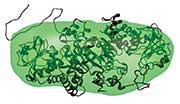
Putting Protein Structure Determination on the Fast Track. Using DOE’s
supercomputing resources and automated small-angle X-ray scattering instrumentation at an
experiment station linked to Berkeley Lab’s Advanced Light Source (right), researchers in
one month resolved the threedimensional structures of 40 proteins from
Pyrococcus
furiosis—a hydrogen-producing microbe thriving in boiling temperatures. One of the
protein structures is pictured at bottom right. This work would have taken several
years with traditional X-ray crystallography techniques. By analyzing the molecular
structures of this heat-tolerant microbe, scientists are working to understand which
protein characteristics enable an organism to withstand the very hot and acidic
conditions associated with biofuel production and other industrial processes.
[Image credits: Lawrence Berkeley National Laboratory]
Pyrococcus
furiosis
Accelerating Macromolecular Structure Determination
with DOE Resources
Synchrotron light sources and neutron facilities at the Department of Energy’s (DOE) national laboratories enable understanding of the structure of matter down to the atomic or molecular level using approaches
not possible with laboratory instrumentation. Synchrotron facilities produce intense beams of photons, from X-rays to infrared to terahertz radiation, while neutron facilities produce beams using particle accelerators or reactors. The beams are directed into experimental stations housing instruments configured for specific
biological investigations.
This infrastructure provides user access to beamlines and instrumentation for high-resolution studies of biological
organisms and molecules for all areas of research in the life sciences. Users are chosen through a peerreviewed
proposal process managed by each facility. Capabilities of and contact information for each station>
are described in the following pages. To find out more about what each experimental program offers, contact
the facilities directly.
This activity is supported by DOE’s Office of Biological and Environmental Research within the Office of Science
and is closely coordinated with other federal agencies and private organizations.
BER's Structural Biology Experimental Stations
- Structural Biology Center at the Advanced Photon Source
The Structural Biology Center (SBC) is a major protein crystallography research facility that enables the atomic-scale study of macromolecular
systems using extremely small (micron-size) crystal samples. SBC’s two experimental stations—the
insertion-device beamline and the bending-magnet beamline—are among the most powerful and focused
X-ray sources available for structural biology. Output is enhanced by on-axis sample viewing optics; easy access
to minibeams (5, 10, and 20 μm) and variable beam sizes (25 to 250 μm); integration of computing and datastorage
resources to accelerate data analysis and archiving; near real-time data interpretation, optimization of
experimental parameters, and structure solution; and full integration of synchrotron hardware, detectors, crystal
mounting robot, beamline software, and crystallographic software packages. These capabilities provide not just
diffraction data, but also an interpretable electron density map and a macromolecular structure.
SBC’s beamlines can be used for a wide range of crystallographic experiments involving:
- Crystals of macromolecular assemblies with very large unit cells
- Multi- or single-wavelength anomalous diffraction
- Crystals of membrane proteins
- Small, weakly diffracting crystals
- Ultra high-resolution crystallography
- Cryo-crystallography
Location: Advanced Photon Source, Argonne National Laboratory, Argonne, IL
Website: http://www.sbc.anl.gov
- PXRR: Macromolecular Crystallography Research Resource at the National Synchrotron Light Source
PXRR provides facilities and support for macromolecular structure determination by synchrotron X-ray diffraction.
Five PXRR beamlines, two of which are high-brightness undulators, allow for highly efficient structure
determination by every available technique. Complementary spectroscopic methods, including optical
absorption spectroscopy and Raman spectroscopy, enable simultaneous measurements of the same sample
under nearly identical experimental conditions. PXRR also provides a popular mail-in crystallography program,
builds new facilities, advances automation, and develops remote data-collection capabilities.
Location: National Synchrotron Light Source, Brookhaven National Laboratory, Brookhaven, NY
Website: http://www.px.nsls.bnl.gov
PDF handout
- Structural Molecular Biology (SMB) Center at the Stanford Synchrotron Radiation Lightsource (SSRL)
The SMB Center’s core areas of technological research and development and scientific focus are macromolecular
crystallography (MC), X-ray absorption spectroscopy (XAS), and small-angle X-ray scattering and diffraction (SAXS).
MC determines the three-dimensional (3-D) structure of biological molecules down to near atomic resolution
(<1 Å), thereby helping elucidate the detailed mechanisms by which macromolecules carry out their functions in
living cells and organisms. MC stations provide high-intensity beams for MAD/SAD and monochromatic data collection
and also offer highly automated robotics-based, high-throughput crystal screening and data collection. All MC
beamlines provide remote access control, allowing users to perform measurements from their home institutions.
A state-of-the-art microbeam station with a high-performance, large-area pixel array detector (PAD) enables study
of the most challenging biomolecular problems (e.g., large macromolecular complexes with large unit cells, small
“micro” crystals, and mechanically and radiation-sensitive samples).
XAS is used to obtain structural information on metal sites in biomolecules. The focus at the optimized XAS beamlines
and instrumentation is on dilute metalloprotein XAS, microbeam imaging/XAS, low-Z XAS (for studies of
ligands such as sulfur and chlorine), and polarized single-crystal XAS studies. The range of XAS equipment includes
advanced solid-state array X-ray fluorescence detector systems, liquid-helium cryostats and Kirkpatrick-Baez optic
micro-XAS instrumentation, and wet-laboratory facilities. The SMB Center provides software for beamline and instrumentation
control, data acquisition, and on- and off-line data analysis.
SAXS features state-of-the-art experimental facilities for solution scattering, lipid membrane diffraction, fiber diffraction,
and single-crystal diffraction at moderately high to very small scattering angles in lengths ranging from microns to a few
angstroms. These techniques allow structural studies of biological macromolecules and assemblies in physiological or
near physiological conditions, complementing high-resolution structural techniques that require experimental conditions
not necessarily physiological. In addition to providing user-friendly experimental facilities for equilibrium studies,
this station maintains premier experimental facilities for time-resolved studies on time scales of milliseconds and above.
Location: Stanford Synchrotron Radiation Lightsource at the SLAC National Accelerator Laboratory, Stanford, CA
Website: http://www-ssrl.slac.stanford.edu/science/smbgroup.html
PDF handout
- Protein Crystallography Station (PCS) at Los Alamos Neutron Science Center
PCS is a high-performance neutron beamline that forms the core of a capability for investigating the structure
and dynamics of proteins, biological polymers, and membranes. Neutron diffraction is a powerful technique
for locating hydrogen atoms, which can be hard to detect using X-rays, and therefore can provide
unique information about how biological macromolecules function and interact with each other and smaller
molecules. PCS users have access to neutron beam time, deuteration facilities, technologies for studying protein
expression and substrate synthesis with stable isotopes, a purification and crystallization laboratory, and software
and support for data reduction and structure analysis. A HomeFlux X-ray system was recently acquired to
allow users to collect X-ray data from the same samples used for neutron diffraction. The PCS beamline exploits
the pulsed nature of spallation neutrons and a large electronic detector to collect wavelength-resolved Laue
patterns using time-of-flight techniques. Data are collected efficiently and with good signal to noise using all
available neutrons [with a wavelength range of ~0.7 to 7.0 angstroms (Å)] in the pulsed white beam.
Location: Los Alamos Neutron Science Center, Los Alamos, NM
Website: http://lansce.lanl.gov/lujan/instruments/PCS.shtml
PDF handout
- Structurally Integrated Biology for the Life Sciences (SIBYLS) at the Advanced Light Source
The SIBYLS beamline is a dual-end station combining macromolecular crystallography (MX) with small-angle
X-ray scattering (SAXS). MX extracts high-resolution structural information from biological molecules, and the
high-throughput SAXS pipeline enables the same biological systems to be imaged in aqueous solution, closer
to their natural state. Combining SAXS results with atomic-resolution structures provides detailed characterizations
in solution of mass, radius, conformations, assembly, and shape changes associated with protein folding and functions.
SAXS also can resolve ambiguities of crystallography by showing the most likely possible structures.
Location: Advanced Light Source, Lawrence Berkeley National Laboratory, Berkeley, CA
Website: http://sibyls.als.lbl.gov/;
See also http://alsusweb.lbl.gov
PDF handout
- Center for Structural Molecular Biology (CSMB) at the High Flux Isotope Reactor and Spallation Neutron Source
CSMB is dedicated to developing instrumentation and methods for determining the structure, function, and
dynamics of complex biological systems. CSMB’s suite of tools includes a small-angle neutron scattering
(SANS) facility for studying biological samples under physiological (or physiologically relevant) conditions,
a bio-deuteration laboratory for in vivo isotopic labeling, and advanced computational resources for modeling
proteins and protein complexes in solution. Deuterium-labeling techniques enable scientists to selectively
highlight and map chemically distinct components of larger protein-protein, protein-lipid, or protein-nucleic
acid complexes and, moreover, to follow their conformational changes and assembly or disassembly processes in
solution on biologically relevant time scales. These capabilities are helping researchers understand how macromolecular
systems are formed and interact with other systems in living cells—ultimately bridging the information
gap between cellular function and the molecular mechanisms that drive it.
Location: Oak Ridge National Laboratory, Oak Ridge, TN
Website: http://www.csmb.ornl.gov
PDF handout
- National Center for X-ray Tomography (NCXT) at the Advanced Light Source
A primary research area at NCXT is development of soft X-ray tomography (SXT), a technique for quantitatively
imaging whole, hydrated cells in 3-D. This technique has several distinct advantages over light and
electron microscopy and is contributing unique insights on cells and their behavior. The soft X-ray illuminating
photons used in SXT penetrate biological materials much more easily than electrons and allow specimens
up to 10 μm thick to be imaged. Unlike electron microscopy, SXT eliminates the need to section eukaryotic cells
with an ultramicrotome before imaging. Since contrast in SXT is produced directly by the differential absorption
of X-rays, specimens do not have to be dehydrated or stained. Consequently, SXT produces high-resolution views
of specimens in a near-native state.
SXT has become an even more powerful technique because of the development of high-aperture cryogenic light
microscopy for correlated imaging. This multimodal approach allows labeled molecules to be localized in the
context of a high-resolution, 3-D tomographic reconstruction of a cell.
Location: Advanced Light Source, Lawrence Berkeley National Laboratory, Berkeley, CA
Website: http://ncxt.lbl.gov/
PDF handout
- Berkeley Synchrotron Infrared Structural Biology (BSISB) Program at the Advanced Light Source
BSISB features synchrotron radiation–based Fourier transform infrared (SR-FTIR) microscopy, which is a label-free,
noninvasive molecular technique that couples the high brightness of synchrotron radiation with the
high throughput and vast analytical capabilities of FTIR spectrometers. With a synchrotron source, BSISB’s
FTIR microscopes are capable of diffraction-limited chemical imaging with signal-to-noise ratios 100 to 1000
times greater than standard blackbody sources.
Aqueous environments hinder SR-FTIR’s sensitivity of bacterial activity, but the recent development of integrated
in situ open-channel microfluidic culturing systems circumvents the water-absorption barrier. These systems
enable real-time chemical imaging of bacterial activities in biofilms and facilitate comprehensive understanding
of structural and functional dynamics in a wide range of microbial systems.
Location: Advanced Light Source, Lawrence Berkeley National Laboratory, Berkeley, CA
Website: http://infrared.als.lbl.gov/
PDF handout
Advanced Biological and Environmental X-Ray Spectroscopy (ABEX) at the Advanced Light Source
ABEX is a new user resource that enables X-ray spectroscopic characterization of complex biological and
environmental systems using soft X-ray absorption (XAS), X-ray magnetic circular dichroism (XMCD), and
resonant inelastic X-ray scattering (RIXS). These spectroscopies exploit the availability of high brightness,
circularly polarized soft X-rays at the Advanced Light Source and offer unique advantages in analyzing the
detailed electronic and magnetic structure of biological metal sites.
ABEX also has a major instrument development program to improve both the sensitivity and user-friendliness of
its instruments. A spectroscopy support laboratory will provide electron paramagnetic resonance (EPR), infrared,
and resonance Raman spectroscopies, enabling essential control measurements on the same samples studied by
the X-ray techniques.
Location: Advanced Light Source, Lawrence Berkeley National Laboratory, Berkeley, CA
Website: http://abex.lbl.gov/
For more information about DOE structural biology resources, contact:
Roland Hirsch
Biological Systems Science Division
U.S. Department of Energy Office of Science
301-903-9009
Email format for contacting Hirsch: firstname.lastname@science.doe.gov


