


What Is Tetralogy of Fallot?
Tetralogy (teh-TRAL-o-je) of Fallot (fah-LO) is a congenital heart defect. This is a problem with the heart's structure that's present at birth. Congenital heart defects change the normal flow of blood through the heart.
Tetralogy of Fallot is a rare, complex heart defect. It occurs in about 5 out of every 10,000 babies. The defect affects boys and girls equally.
To understand tetralogy of Fallot, it helps to know how a healthy heart works. The Health Topics How the Heart Works article describes the structure and function of a healthy heart. The article also has animations that show how your heart pumps blood and how your heart's electrical system works.
Overview
Tetralogy of Fallot involves four heart defects:
- A large ventricular septal defect (VSD)
- Pulmonary (PULL-mun-ary) stenosis
- Right ventricular hypertrophy (hi-PER-tro-fe)
- An overriding aorta
Ventricular Septal Defect
The heart has an inner wall that separates the two chambers on its left side from the two chambers on its right side. This wall is called a septum. The septum prevents blood from mixing between the two sides of the heart.
A VSD is a hole in the septum between the heart's two lower chambers, the ventricles. The hole allows oxygen-rich blood from the left ventricle to mix with oxygen-poor blood from the right ventricle.
Pulmonary Stenosis
This defect involves narrowing of the pulmonary valve and the passage from the right ventricle to the pulmonary artery.
Normally, oxygen-poor blood from the right ventricle flows through the pulmonary valve and into the pulmonary artery. From there, the blood travels to the lungs to pick up oxygen.
In pulmonary stenosis, the pulmonary valve cannot fully open. Thus, the heart has to work harder to pump blood through the valve. As a result, not enough blood reaches the lungs.
Right Ventricular Hypertrophy
With this defect, the muscle of the right ventricle is thicker than usual. This occurs because the heart has to work harder than normal to move blood through the narrowed pulmonary valve.
Overriding Aorta
This defect occurs in the aorta, the main artery that carries oxygen-rich blood from the heart to the body. In a healthy heart, the aorta is attached to the left ventricle. This allows only oxygen-rich blood to flow to the body.
In tetralogy of Fallot, the aorta is located between the left and right ventricles, directly over the VSD. As a result, oxygen-poor blood from the right ventricle flows directly into the aorta instead of into the pulmonary artery.
Outlook
With tetralogy of Fallot, not enough blood is able to reach the lungs to get oxygen, and oxygen-poor blood flows to the body.
Cross-Section of a Normal Heart and a Heart With Tetralogy of Fallot
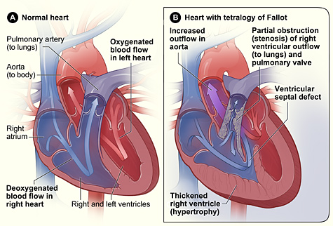
Figure A shows the structure and blood flow inside a normal heart. Figure B shows a heart with the four defects of tetralogy of Fallot.
Babies and children who have tetralogy of Fallot have episodes of cyanosis (si-ah-NO-sis). Cyanosis is a bluish tint to the skin, lips, and fingernails. It occurs because the oxygen level in the blood leaving the heart is below normal.
Tetralogy of Fallot is repaired with open-heart surgery, either soon after birth or later in infancy. The timing of the surgery will depend on how narrow the pulmonary artery is.
Over the past few decades, the diagnosis and treatment of tetralogy of Fallot have greatly improved. Most children who have this heart defect survive to adulthood. However, they'll need lifelong medical care from specialists to help them stay as healthy as possible.

Clinical trials are research studies that explore whether a medical strategy, treatment, or device is safe and effective for humans. To find clinical trials that are currently underway for Tetralogy of Fallot, visit www.clinicaltrials.gov.
![]()
Visit Children and Clinical Studies to hear experts, parents, and children talk about their experiences with clinical research
The NHLBI updates Health Topics articles on a biennial cycle based on a thorough review of research findings and new literature. The articles also are updated as needed if important new research is published. The date on each Health Topics article reflects when the content was originally posted or last revised.