
Printer-friendly version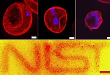
A joint research team, working at Commerce’s National Institute of Standards and Technology (NIST) and the National Institute of Allergy and Infectious Diseases (NIAID), has discovered a method of using nanoparticles to illuminate the cellular interior to reveal these slow processes. Nanoparticles, thousands of times smaller than a cell, have a variety of applications. One type of nanoparticle called a quantum dot glows when exposed to light. These semiconductor particles can be coated with organic materials, which are tailored to be attracted to specific proteins within the part of a cell a scientist wishes to examine. (More)
Comments Closed
Due to increased spam, comments have been closed on this content. If you wish to comment about the content, we encourage you to email webmaster@doc.gov.