 |
 |
| Malaria
Home > |
|
A Practical Tool for the Microscopic Diagnosis of Malaria
|
|
 |
During a malaria survey in the Democratic Republic of Congo, health workers identify infected persons by examining their blood smears under the microscope.
(Courtesy Ministry of Health, Democratic Republic of Congo) |
When a person is suspected of having malaria, the diagnosis should be confirmed before the patient is treated. To do this, health workers most often:
- take a small blood sample from the patient, most of the time through a fingerprick
- spread this specimen on a glass slide, making thick and thin blood smears
- stain the blood smears in a special stain (Giemsa’s)
- examine the stained blood smears under a microscope.
If malaria is present, the microscopic examination will show parasites infecting some of the patient’s red blood cells.
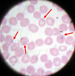 |
Blood smear from a patient with malaria; microscopic examination shows Plasmodium falciparum parasites (arrows) infecting some of the patient's red blood cells. (CDC photo) |
To examine a specimen under the microscope, a strong light beam must be shined on the specimen. This is usually achieved with a light bulb incorporated in the microscope. However, in remote areas of poor countries, where most of malaria cases worldwide occur, electric power may be absent or unreliable. Under these circumstances, laboratory workers use a mirror mounted on the microscope to reflect sunlight onto the specimen. This method however cannot be used at night or during cloudy or rainy days.
A practical solution to this problem is now available. Working in collaboration at CDC, Dr. Earl Long, a research microbiologist, and Mr. Charles Boozer, a biomedical engineer, developed a portable, battery-operated white light-emitting diode that can be placed next to a microscope. This “EARL-Light” provides excellent illumination for up to 1,250 hours, thus allowing microscopy in the absence of electricity and sunlight.
Click for EARL-Light Power Point Presentation.
 |
Mr. Jean-Pierre Masendu putting the final touch in the manufacture of an EARL-Light in Kinshasa, Democratic Republic of Congo. (Courtesy Ministry of Health, Democratic Republic of Congo) |
After successful field trials, the EARL-Light is now used in several countries for the microscopic diagnosis of malaria as well as other infectious diseases, such as tuberculosis.
 |
A laboratory worker in Tanzania shines the light beam of an EARL-Light onto a mirror, which reflects it on the specimen being examined under the microscope; the device thus replaces sunlight. (Courtesy CDC/Ifakara Health Research and Development Centre Malaria Programme) |
Page last modified : August 24, 2006
Content source: Division of Parasitic Diseases
National Center for Zoonotic, Vector-Borne, and Enteric Diseases (ZVED)
|
|
|