| Newsroom |
Split beamlines can double research capacity at Advanced Photon SourceNew structural biology beamline is first to use split beamlines to double research capacity and to create finer X-ray beams to capture data from tough-to-study biomoleculesARGONNE, Ill. (Aug. 19, 2005) — A new beamline dedicated this summer at the Advanced Photon Source (APS) sets a new standard for structural biology research at synchrotrons. The GM/CA CAT facility exploits the latest technology to double the number of beamlines and create finer X-ray beams to capture data from hard-to-study biomolecules.
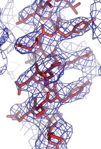
This development comes at a time of growing need for beamlines. Structural biologists use X-ray beamlines in a technique called protein crystallography to study the three-dimensional structures of molecules critical to life. Understanding the structure and functions of materials that control muscle motion or cause influenza, for example, allows researchers to create new drugs to treat, cure or prevent diseases. The new GM/CA CAT facility is a collaboration between the U.S. Department of Energy and the National Institutes of Health's National Institute for General Medical Sciences (GM) and National Cancer Institute (CA). Three new beamlines will be available at the APS – the source of the Western Hemisphere 's most brilliant X-ray beams – by January 2007. “The need for structural biology beam time continues to grow,” said Janet Smith, GM/CA CAT director, “and this facility will help provide researchers from the NIH and other organizations the tools to discover how proteins and biomolecules work.” Smith, a structural biologist at the University of Michigan in Ann Arbor, directs a team of 13 Argonne Biosciences Division employees. Getting to the sourceBefore a protein or biomolecule makes it to a synchrotron beamline, it must be produced from DNA in a cell, purified, crystallized and frozen. When X-rays pass through the crystal, they are diffracted as thousands of scattered X-ray beams are recorded in hundreds of two-dimensional images. Crystallographers analyze the diffraction images to produce a three-dimensional image of the molecule in the crystal. The results are published and contributed to the Protein Data Bank, the international data base for protein structures. Biologists search this data base for structural patterns, for example, to deduce how proteins fold in a particular shape or to discover clues to how a drug might bind to a protein and affect its function. In 2004, researchers using the APS led the world in Protein Data Bank deposits. Best biology beamlinesThe GM/CA CAT team took advantage of the best available technologies to build a state-of-the-art facility. The beamline optics were designed together with ACCEL Instruments, GmbH, the contractor who fabricated and installed the components." The facility has a number of claims to fame:
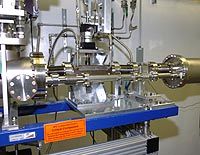
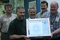
The APS circulates electrons in an enclosed racetrack. The electrons are manipulated by undulators magnetic devices that produce intense X-ray beams. The X-ray beams exit the racetrack through ports into each of the beamlines. For the GM/CA beamlines, the APS installed two undulators instead of one – to produce two X-ray beams of equal intensity in slightly different directions – just a few hair-widths apart at the source. The X-ray beams separate as they travel about 60 meters to the first experimental station, where they are about two feet apart. One beam passes through to the second experimental station, about 75 meters from the source. Experiments can be run in the two stations simultaneously. “The ability to do two experiments simultaneously from the same light source is a big bonus,” said Smith. “It doubles the value of the real estate at the synchrotron.” The GM/CA beamlines also boast the world's best large X-ray mirrors. “The mirrors have to be properly shaped in order to focus the X-rays,” explained GM/CA CAT Project Manager Bob Fischetti. “All mirrors appear smooth to the human eye. We need the mirrors to appear smooth to X-rays because smooth mirrors provide a small, clean beam, and the result is more precise data.” These mirrors are built by sandwiching small piezoelectric ceramic plates between silica plates. The ceramic plates can be bent by applying a voltage to electrode patches on them. By applying the appropriate voltages to the many electrodes, the mirror can be perfectly shaped. SESO, the French company that made the mirrors, pushed its technology to make the GM/CA CAT beamline mirrors more than a meter long. Previously these “bimorph” mirrors were only about 300 millimeters long. Beam-position monitors keep the X-ray beam in place during its 60-to-75-meter jaunt to the sample by providing feedback that keeps the X-rays on target. The GM/CA CAT team placed a beam-position monitor directly in front of the experimental sample, and the world's smoothest mirrors tweak the beam into its precise position. “This narrow beam,” Smith said, “allows us to irradiate only the crystal – not the crystal surroundings – or just the part of the crystal we wish to study. This is important because anything else in the X-ray beam scatters X-rays and adds noise to the diffraction pattern. If we can reduce the noise, we can detect weaker signals and study smaller crystals.” The advanced optics allows researchers to study both smaller crystals and larger molecules than before. Some protein crystals are just smaller than others, Fischetti said, “and smaller crystals are easier to make and may be higher quality.” On the other hand, a virus particle is huge, so that virus crystals produce hundreds of thousands of diffracted X-ray beams that are very close together. Early resultsThe beamline has already produced valuable data for publication by researchers at the Scripps Research Institute. Lu Gan and colleagues studied a crystal of the complex virus HK97 as it underwent a staged assembly process where protein subunits combine. By using the narrow beam to avoid overlapping diffraction peaks and to enhance the signal above noise, Gan was able “to position all of the side chains and to establish a full chemical understanding of the stabilizing forces of the expansion structure he was studying,” said Jack Johnson of Scripps, who chairs the GM/CA CAT advisory board. “Previously we were only able to follow the polypeptide chain of the proteins with little confidence in the side chain positions.” Some other large biological systems that the GM/CA CAT can study include large multi-protein complexes such as myosin which controls muscle motion, ribosome which reads the genetic code to produce proteins, ferritin which controls iron storage and transport in the body, and chaperonins which facilitate protein folding in cells. Easy operations “One of the goals of this project was to design experimental facilities that are robust and easy to operate, allowing users to focus on their samples instead of which buttons to push,” said Smith. “Our on-axis visualization system allows researchers to easily align the X-ray beam to the sample,” Fischetti said. Previously, users had to use cameras at inconvenient angles or mirrors to align these very small samples. On-axis visualization saves the researcher time and improves the data quality. Johnson concurs. “The user interface on these beam lines is based on a system called ‘Blu-Ice' used on other advanced crystallography beam lines. It was relatively straightforward to implement this comprehensive and familiar graphical interface for controlling the beamline.” The GM/CA CAT also will use a sample-handling robot. “Our samples are typically cryo-preserved in liquid nitrogen,” Smith said. “The robot will take the frozen samples from a liquid nitrogen storage unit and put them on the diffraction instrument, all the while keeping them cold.” Changing times for synchrotrons and structural biology “The Advanced Photon Source is becoming the international powerhouse of structural biology,” said Biosciences Division Director Lee Makowski. The APS provides significant contributions of protein structures to the Protein Data Bank. In 2004, the APS contributed 21 percent of all of the X-ray structures from synchrotron facilities, according to the Protein Data Bank. The APS is also home to ID-19, the world's most efficient structural biology beamline. Today ID-19 provides more structures to the Protein Data Bank than any other beamline in the world. When the first GM/CA CAT beamline begins full production mode in January 2006, it will set a new standard for structural biology beamlines. It will double or triple the structure output of ID-19, according to Andzrej Joachimiak, the director of the Structural Biology Center that operates ID-19. The second of the GM/CA CAT dual-canted undulator beamlines that uses the same technologies as the first, is currently being commissioned. The third beamline is based on a bending magnet source with conventional X-ray mirrors, but will otherwise use the same technologies as the undulator counter parts, which will be in full production mode by June 2007. In addition, two other structural biology CATs using the dual-canted undulator technology are under construction at the APS. “The APS has more structural biology beamlines than any other third-generation synchrotron source,” Makowski said. “Its impact in biology will only continue to grow.” — Evelyn Brown For more information, please contact Steve McGregor (630/252-5580 or media@anl.gov) at Argonne. |
Resources |
||
|
| U.S. Department of Energy Office of Science | UChicago Argonne LLC |
| Privacy & Security Notice | Contact Us | Site Map | Search |