What Is Tetralogy of Fallot?
Tetralogy (teh-TRAL-o-je) of Fallot (fah-LO) is a
congenital
heart defect. A congenital heart defect is a problem with the heart's
structure that’s present at birth. This type of heart defect changes the
normal flow of blood through the heart.
Tetralogy of Fallot is a rare, complex heart defect
that occurs in about 5 out of every 10,000 babies. It affects boys and
girls equally.
To understand this defect, it's helpful to know how
a healthy heart works. The Diseases and Conditions Index
How
the Heart Works article describes the structure and function of a healthy
heart. The article also has animations that show how your heart pumps blood and
how your heart's electrical system works.
Overview
Tetralogy of Fallot involves four heart defects:
- A large
ventricular
septal defect (VSD)
- Pulmonary (PULL-mon-ary) stenosis
- Right ventricular hypertrophy (hi-PER-tro-fe)
- An overriding aorta
Ventricular Septal Defect
The heart has a wall that separates the two chambers
on its left side from the two chambers on its right side. This wall is called a
septum. The septum prevents blood from mixing between the two sides of the
heart.
A VSD is a hole in the part of the septum that
separates the ventricles, the lower chambers of the heart. The hole allows
oxygen-rich blood from the left ventricle to mix with oxygen-poor blood from
the right ventricle.
Pulmonary Stenosis
This defect is a narrowing of the pulmonary valve
and the passage through which blood flows from the right ventricle to the
pulmonary artery.
Normally, oxygen-poor blood from the right ventricle
flows through the pulmonary valve, into the pulmonary artery, and out to the
lungs to pick up oxygen. In pulmonary stenosis, the heart has to work harder
than normal to pump blood, and not enough blood reaches the lungs.
Right Ventricular Hypertrophy
This defect occurs if the right ventricle thickens
because the heart has to pump harder than it should to move blood through the
narrowed pulmonary valve.
Overriding Aorta
This is a defect in the aorta, the main artery that
carries oxygen-rich blood to the body. In a healthy heart, the aorta is
attached to the left ventricle. This allows only oxygen-rich blood to flow to
the body.
In tetralogy of Fallot, the aorta is between the
left and right ventricles, directly over the VSD. As a result, oxygen-poor
blood from the right ventricle flows directly into the aorta instead of into
the pulmonary artery to the lungs.
Outlook
Together, these four defects mean that not enough
blood is able to reach the lungs to get oxygen, and oxygen-poor blood flows out
to the body.
Normal Heart and Heart With
Tetralogy of Fallot
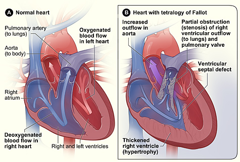
Figure A shows the structure and
blood flow in the interior of a normal heart. Figure B shows a heart with the
four defects of tetralogy of Fallot.
Babies and children who have tetralogy of Fallot
have episodes of cyanosis (si-a-NO-sis). This is a bluish tint to the skin,
lips, and fingernails. Cyanosis occurs because the oxygen level in the blood is
below normal.
Tetralogy of Fallot must be repaired with open-heart
surgery, either soon after birth or later in infancy. The timing of the surgery
depends on how severely the pulmonary valve is narrowed.
Over the past few decades, the diagnosis and
treatment of tetralogy of Fallot have greatly improved. As a result, most
children who have this heart defect survive to adulthood. However, they’ll
need lifelong medical care from specialists to help them stay as healthy as
possible.
Revised August 2009 |