
![]()
Home
About CFCF
Board of Directors
Board of Patient Advisors
Board of Scientific Advisors
Carcinoid Community
Carcinoid Information
Clinical Trials
Donations
Friends of CFCF
Fundraising Events
How You Can Help
Memorials
News
Photo Gallery
Research
Sponsors
Sunflower
About Carcinoid
Overview
Carcinoid Definition
Carcinoid is a rare, deadly neuroendocrine cancer. The American Cancer Society defines carcinoid as the following:
"Like most cells of the body, gastrointestinal system neuroendocrine cells sometimes undergo certain changes that cause them to grow too much and form tumors. The tumors that develop from neuroendocrine cells are known as neuroendocrine tumors (or neuroendocrine cancers). There are many varieties of neuroendocrine tumors, but the most common are the carcinoid tumors or carcinoids.
Carcinoid tumors act like the cells they come from. They often release certain hormone-like substances into the bloodstream. In about 10% of people, the carcinoid tumors spread and grow very large and release high amounts of those hormones. These cause symptoms such as facial flushing (redness and warm feeling), wheezing, diarrhea, and a fast heartbeat. These symptoms are grouped together and called the "carcinoid syndrome." Most cancers cause symptoms only in the organs they start in or spread to. But carcinoid tumors can release substances into the blood that cause symptoms throughout the body."
Carcinoid Types and Locations
Carcinoid Types: Foregut, Midgut, and Hindgut - Novartis (video)
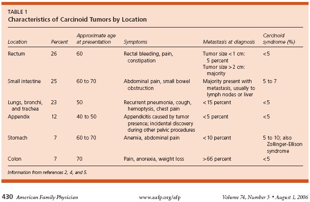
Carcinoid Characteristics by Location - American Family Physician


Carcinoid Genetics
Genetics of Neuroendocrine and Carcinoid Tumors - Endocrine-Related Cancer


Carcinoid Presentations
Carcinoid Tumors: Current Update on Tumor Biology, Diagnosis and Therapy - Dr. Kjell Oberg

Carcinoid Guides
Guide to Carcinoid Tumor - People Living with Cancer


Neuroendocrine Tumors - Dana-Farber Cancer Institute


Carcinoid Articles
Metastatic Carcinoid Tumors: A Clinical Review - The Oncologist
Carcinoid Tumors - American Family Physician
Carcinoid Tumors - Endotext.org
Carcinoid Syndrome - MedicineNet.com
Gastrointestinal Carcinoid Tumor - American Cancer Society


Causes
Very little is known about the causes of carcinoid, according to the American Cancer Society:
"Very little is known about the causes of gastrointestinal carcinoid tumors.
As with other cancers, scientists have recognized some changes in the DNA of carcinoid tumor cells that are probably responsible for their increased growth and abnormal spread. But the causes of these changes are not yet known.
Doctors do know that carcinoid tumors start out very small and grow slowly. When patients have parts of their stomach or small intestine removed to treat other diseases, close examination under the microscope often shows small groups of neuroendocrine cells that look like tiny carcinoids. Most of these miniature tumors, sometimes called tumorlets, never develop into an actual carcinoid tumor. Researchers still do not know why some remain so small and others begin to grow and become large enough to cause symptoms."
Diagnosis and Testing
Introduction
The following articles provide an introduction to diagnosis and testing of carcinoid:
Diagnosis and Management of Neuroendocrine Tumors - Medscape
Diagnosis and Medical Management of Advanced Neuroendocrine Tumors - Endocrine Reviews
The following questionnaire may be helpful for your physician if you think you have carcinoid

5-HIAA
The following information is provided by the American Cancer Society:
"Serotonin is one of the substances produced by some carcinoid tumors, especially those developing in the small intestine. Serotonin in the blood can be measured. Also, it is broken down to 5-hydroxyindoleactic acid (abbreviated 5-HIAA), which is released into the urine. Urine tests to measure 5-HIAA levels are very useful in diagnosing carcinoid tumors that produce serotonin and have spread to the liver. However, localized gastrointestinal carcinoid tumors often do not have positive urine 5-HIAA results."
5-HIAA Test - Lab Tests Online
Foods and Drugs to Avoid When Performing 5-HIAA Test - Cleveland Clinic


Barium X-rays
The following information is provided by the American Cancer Society:
"These x-ray studies use a barium-containing solution that coats the lining of the esophagus, stomach, and intestines. These are often useful for the diagnosis of some gastrointestinal carcinoid tumors. They are least effective in finding small intestine carcinoid tumors. The coating of barium helps find abnormalities of the lining of these organs. Barium studies can be used to examine the upper or lower parts of the digestive system."
Biopsy
The following information is provided by the American Cancer Society:
"Even if a barium x-ray and/or CT scan finds a mass, these imaging tests cannot tell if the mass is a carcinoid tumor, some other type of tumor (benign or cancerous), or a localized infection. The only way to know for sure is to remove cells from the abnormal area and examine them under a microscope. This procedure is called a biopsy."

Chromogranin A
The following information is provided by the American Cancer Society:
"Blood tests may be done to detect some of the hormone-like substances produced by carcinoid tumors, particularly if the patient has symptoms of the carcinoid syndrome, caused by excessive levels of such substances in the blood. The most commonly used blood tests measure levels of chromogranin A or neuron-specific enolase. Depending on the patient's symptoms, doctors may recommend additional blood tests."
Chromogranin A Test - Novartis
The Chromogranin–Secretogranin Family - New England Journal of Medicine


Colonoscopy
The following information is provided by the National Institute of Diabetes and Digestive and Kidney Diseases:
"A colonoscopy allows a doctor to look inside the entire large intestine. The procedure enables the physician to see things such as inflamed tissue, abnormal growths, and ulcers. It is most often used to look for early signs of cancer in the colon and rectum. It is also used to look for causes of unexplained changes in bowel habits and to evaluate symptoms like abdominal pain, rectal bleeding, and weight loss."
Computed Tomography (CT)
The following information is provided by the American Cancer Society:
"The CT scan is an x-ray procedure that produces detailed cross-sectional images of your body. Instead of taking one picture, like a conventional x-ray, a CT scanner takes many pictures as it rotates around you. A computer then combines these pictures into an image of a slice of your body. The machine takes pictures of multiple slices of the part of your body that is being studied. This test can help tell if your carcinoid tumor has spread into lymph nodes or other organs such as your liver. Often after the first set of pictures is taken you will receive an intravenous (IV) injection of a contrast agent, or dye, which helps better outline structures in your body. A second set of pictures is then taken.
CT scans can also be used to guide a biopsy needle precisely into a suspected metastasis. For this procedure, called a CT-guided needle biopsy, the patient remains on the CT scanning table, while a radiologist moves a biopsy needle toward the location of the mass. CT scans are repeated until the doctors are confident that the needle is within the mass. A fine-needle biopsy sample (tiny fragment of tissue) or a core-needle biopsy sample (a thin cylinder of tissue about ½-inch long and less than 1/8-inch in diameter) is removed and examined under a microscope.
CT scans are more tedious than regular x-rays because they take longer and you need to lie still on a table while they are being done. But just like other computerized devices, they are getting faster and your stay might be pleasantly short. Also, you might feel a bit confined by the ring you lay within when the pictures are being taken.
You will need an IV line through which the contrast dye is injected. The injection can cause some flushing. Some people are allergic and get hives, or rarely, more serious reactions like trouble breathing and low blood pressure can occur. Please be sure to tell the doctor if you have ever had a reaction to any contrast material used for x-rays."

Endoscopy
The following information is provided by the American Cancer Society:
"This test uses a flexible lighted tube with a video camera on the end. The camera is connected to a monitor, allowing the doctor to clearly see any masses in the lining of the digestive organs. If abnormalities are noted, small pieces of tissue can be removed through the endoscope (biopsy). The tissue can be examined under the microscope to find out if cancer is present and what kind of cancer it is."
Endoscopic Ultrasound
The following information is provided by the American Cancer Society:
"This is a new technique in which a special instrument is used in patients having endoscopy. For this test, the endoscope has a small ultrasound probe on the end. This probe releases high frequency sound waves and then detects the sound wave echoes that bounce off tissues of the stomach wall. A computer then translates the pattern of echoes into an image of the wall of the esophagus, stomach, intestine, or rectum.
Endoscopic ultrasound is sometimes useful in determining how far a tumor has spread through the wall of the esophagus, stomach, intestine, or rectum. The test can also help predict whether the tumor has spread beyond the wall of these organs to nearby tissues or lymph nodes."
Positron Emission Tomography (PET)
The following information is provided by the American Cancer Society:
"This is a special kind of radioactive scan. PET scanning for carcinoid tumors uses a radioactive form of 5-hydroxytryptophan, a chemical that is taken up and used by carcinoid cells. A special camera can detect the radioactivity. PET scans are useful when your doctor thinks the cancer has spread but doesn't know where. PET scans can be used instead of several different x-rays because it scans your whole body. Some doctors have found it to be more accurate than a CT scan for detecting spread of disease."
Imaging of Advanced Neuroendocrine Tumors with 18F-FDOPA PET - Journal of Nuclear Medicine
PET in the Diagnosis of Neuroendocrine Tumors - Annals of the New York Academy of Sciences (abstract)

Radioactive Scans
The following information is provided by the American Cancer Society:
"Two procedures have been used. The older one is called I131-MIBG scan. This procedure uses a chemical called MIBG to which radioactive iodine (I131) is attached. This is injected into your vein and then your body is scanned to look for areas that picked up the radioactivity. These would most likely be carcinoid tumors, although other kinds of neuroendocrine tumors will also pick up this chemical.
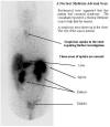
A second kind of scan is indium111-labeled DTPA-octreotide scintigraphy or Octreoscan. Octreotide is a hormone-like substance that attaches to carcinoid cells. A small amount of this radioactive octreotide is injected into a vein. This material is attracted to carcinoid tumors. A special camera scans your body to show where the radioactivity collects. This procedure and the I131-MIBG scan are useful in detecting spread of gastrointestinal carcinoid tumors to other areas of the body."
Octreoscan of Carcinoid - Ninewells Hospital
What is an Octreoscan? - Mid-South Imaging
Scintigraphic Evaluation of Neuroendocrine Tumors - Applied Radiology


Sponsor
CFCF is grateful to Covidien for sponsoring the Diagnostics Section of our website.

Stages
Carcinoid stages indicate how local or widespread is the disease, according to the American Cancer Society:
"Staging – or determining the stage of disease – is the process of finding out how localized or widespread the carcinoid tumor is. It will show whether the tumor has spread and how far. The treatment and prognosis (the outlook for chances of survival) for a patient with a gastrointestinal carcinoid tumor depends, to a large extent, on the tumor's stage.
There is no standard system for describing the spread of gastrointestinal carcinoid tumors. Some doctors use the same systems that are used for other cancers of the same organs. Many doctors simply divide all gastrointestinal carcinoid tumors into 3 general stages: localized, regional spread, and distant spread. This approach is easy for patients to understand and is useful in considering treatment options:
- Localized: The carcinoid tumor has not spread beyond the wall of the organ it developed in (for example, the stomach, intestine, or rectum).
- Regional spread: The carcinoid tumor has spread through the wall of the organ it started in to involve nearby tissues such as fat, ligaments, muscle, or lymph nodes.
- Distant spread: The carcinoid tumor has spread to tissues or organs that are not near the organ where the cancer started. Spread of a gastrointestinal carcinoid tumor to the liver, bones, or lungs, for example, is considered distant spread."
Statistics
Carcinoid statistics, according to the American Cancer Society, are the following:
- 11,000 to 12,000 carcinoid tumors are diagnosed each year in the United States
- The number of carcinoid tumors diagnosed each year has been increasing around 6% per year
- Carcinoid tumors are slightly more common in females than males
- Approximately 50% of carcinoid tumors occur in the digestive system, 30% in the lungs, and 20% in other organs
Updated Population-Based Review of Carcinoid Tumors - Annals of Surgery
A 5-Decade Analysis of 13,715 Carcinoid Tumors - American Cancer Society
Symptoms
The symptoms of carcinoid, according to Novartis, are the following:
Arthritis
"Although the causes are not known, people with carcinoid syndrome are more likely than the general population to develop arthritis."
Carcinoid Heart Disease
"One of the more serious symptoms of carcinoid syndrome is heart valvular lesions, a condition in which excess serotonin causes injury to the valves of the heart. This leads to a unique set of problems with the way your heart functions, called carcinoid heart disease. A little more than half of people with carcinoid syndrome develop heart valvular lesions."
Carcinoid Heart Disease - Novartis (video)
Carcinoid Heart Disease: Presentation, Diagnosis, and Management - Heart
Factors Associated with Progression of Carcinoid Heart Disease - New England Journal of Medicine


Cramping
"About half of people with carcinoid syndrome experience abdominal cramping, a painful condition in which normal bowel movements are prevented. These cramping episodes may occasionally develop into intestinal obstruction, a serious condition that requires medical attention."
Cyanosis
"Cyanosis refers to characteristic bluish skin spots that develop in about 1 in 5 people with carcinoid syndrome. The spots may appear after flushing, and are produced by lack of oxygenated blood circulation in the affected areas."
Diarrhea
"Over 75% of people with carcinoid syndrome experience diarrhea, which can occur with flushing or by itself. Stools are watery and the diarrhea can be mild or severe. Episodes can occur several times a day and can interfere with daily life. Patients with severe cases of diarrhea often have trouble leaving their homes for work, social functions, or activities that require being away from home and on the move for a long time. In addition, diarrhea can drain your body of water, causing dehydration and electrolyte loss. These electrolytes, such as sodium and potassium, help carry electrical charges or messages throughout your body. Without enough water and electrolytes, proper digestion cannot occur, and your body cannot get the nutrients it needs. This can worsen the weight loss, weakness, and fatigue that may have already have been caused by the loss of fluids and electrolytes."
Diarrhea - Novartis (video)

Flushing
"Over 90% of people with carcinoid syndrome experience flushing. Flushing resembles an intense blush, a deep red or purple hue that appears suddenly on the face or neck — although the flush may appear on the trunk, back, or legs as well. The flush can be triggered by emotions, by eating, or by drinking alcohol or hot liquids. When it occurs, you may feel warm or unpleasant sensations in the affected areas, and be aware of having a rapid heartbeat. The flush is caused by dilation of the blood vessels in the affected area of skin. It can last from a few minutes to hours, and in some cases may even be constant. The stage of carcinoid syndrome you are in can determine how long flushing lasts. In more severe cases, facial skin sometimes thickens and discolors."
Flushing - Novartis (video)
Factors that Precipitate Flushing - Cleveland Clinic


Pellagra (Niacin deficiency)
"Pellagra is a disease of nutritional deficiency that causes symptoms such as skin rash because of the lack of niacin."
Peripheral Edema
"About 1 in 5 people with carcinoid syndrome has peripheral edema, a swelling of the ankles, legs, hands and arms, or neck and face. This symptom may be a sign of heart problems, and you should see your doctor if you notice it."
Telangiectasia
"Over a quarter of people with carcinoid syndrome have telangiectasia, reddish spots or veins that appear most often on the face, chest or arms. These are caused by prolonged flushing."
Wheezing
"Exposure of lung tissues to abnormally high levels of certain substances can cause the blood vessels to constrict, and narrow the airway passages, making it difficult to breathe. This wheezing can be mistaken for asthma. About 1 in 5 people with carcinoid syndrome experience this symptom."
Wheezing - Novartis (video)


Dr Aaron Vinik is the doctor on call for April. Click
here to submit your
questions on biomarkers in neuroendocrine tumors and other endocrinology
questions you have regarding NETs.
CFCF Profile


![]()
![]()
CFCF Headlines
Nancy O'Hagan Writes New Blog
The Great Race of Agoura Hills
Seattle to Portland Bicycle Classic

Is it possible to cure carcinoid?
How long will it take to
cure carcinoid?
How much money will be needed to cure carcinoid?
Will achieving our mission benefit all carcinoid patients?