All Images

Press Release 09-088
Shift in Simulation Superiority

New report highlights strengths and weaknesses in U.S. high-end computer simulations relative to international counterparts
Back to article | Note about images
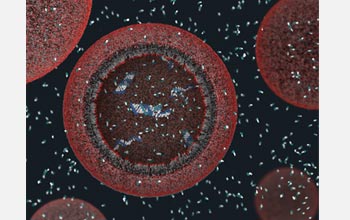 |
Above is a three-dimensional view of a model protocell approximately 100 nanometers in diameter. The protocell's fatty acid membrane allows nutrients and DNA building blocks to enter the cell and participate in non-enzymatic copying of the cell's DNA. The newly formed strands of DNA remain in the protocell. For more about the computer simulations behind this work, including video and animations, see the press release "A New Way to Think About Earth's First Cells".
Credit: Janet Iwasa, Szostak Laboratory, Harvard Medical School and Massachusetts General Hospital |
Download the high-resolution JPG version of the image. (7 MB)
|
Use your mouse to right-click (Mac users may need to Ctrl-click) the link above and choose the option that will save the file or target to your computer.
|
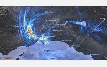 |
This visualization illustrates some of the rupture and wave propagation phenomena of a magnitude 7.8 earthquake on the San Andreas fault in southern California ('the BIG ONE'). The animation captures more than 4 minutes of the complex dynamic rupture and wave propagation for such a large event, including directivity effects, wave-guide phenomena, and local amplification of the ground shaking. The visualization presents these physical effects in an intuitive yet accessible manner using a variety of visual representations and several views. 11.8 TB of earthquake simulation output from a state-of-the-art earthquake simulation was used to generate the animation. Read a press release from ORNL about the work and view the animation.
This animation shows the sliprate on the 16 km deep vertical plane along the southern San Andreas fault and the horizontal plane shows the velocity magnitude on the surface. The animation is based on the TeraShake 2.1 simulations.
Credit: Visualization: Amit Chourasia (SDSC/UCSD)
Simulation: Kim Olsen and Steven Day (San Diego State University); Luis Dalguer (ETH-Zurich); Yifeng Cui and Jing Zhu (SDSC/UCSD); David Okaya, Phil Maechling and Thomas H. Jordan (University of Southern California)
San Diego Supercomputer Center (SDSC), Southern California Earthquake Center (SCEC)
Acknowledgements: Mahidhar Tatineni (SDSC/UCSD), Geoffrey Ely (SCEC), Krishna Muriki (SDSC/UCSD), Eva Hocks (SDSC/UCSD), Douglas Weimer (SDSC/UCSD), Tommy Minyard (TACC), Karl Schulz (TACC), Dongju Choi (SDSC/UCSD), Tim Kaiser (SDSC/UCSD), Sweta Gupta (SDSC/UCSD) |
Download the high-resolution JPG version of the image. (6.7 MB)
|
Use your mouse to right-click (Mac users may need to Ctrl-click) the link above and choose the option that will save the file or target to your computer.
|
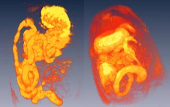 |
Three-dimensional reconstructions of magnetic resonance images of the rat gastrointestinal tract, from the stomach (top) through the small intestines (duodenum, jejunum and caecum). For more information, read the NSF Discovery "Gut Reaction: Digestion Revealed in 3-D."
Credit: Thomas Neuberger and Andrew G. Webb , Huck Institute Magnetic Resonance Centre, Pennsylvania State University |
Download the high-resolution JPG version of the image. (67 KB)
|
Use your mouse to right-click (Mac users may need to Ctrl-click) the link above and choose the option that will save the file or target to your computer.
|
|

