What Is Chest MRI?
Chest magnetic resonance imaging (MRI) is a safe,
noninvasive test. “Noninvasive” means that no surgery is done and
no instruments are inserted into your body. This test creates detailed pictures
of the structures in your chest, like your chest wall, heart, and blood
vessels.
Chest MRI uses radio waves, magnets, and a computer
to create these pictures. The test is used to:
- Look for tumors in the chest
- Look at blood vessels, lymph (limf) nodes, and
other structures in the chest
- Help explain results of other tests, such as
chest
x ray or chest CT scan (also called
computed tomography (to-MOG-rah-fee) scans)
As part of some chest MRIs, a special substance
(called contrast dye) is injected into a vein in your arm. This dye allows the
MRI to take more detailed pictures of the structures in your chest.
Chest MRI has few risks. Unlike a CT scan or
standard x ray, MRI doesn’t use radiation or have any risk of causing
cancer. Rarely, the contrast dye used for some chest MRIs may cause an allergic
reaction.
Other Names for Chest MRI
Chest MRI also may be called chest nuclear magnetic
resonance.
Who Needs a Chest MRI?
You may need a chest MRI if your doctor suspects you
have a chest condition, such as:
- A tumor
- Problems in the blood vessels, such as an
aneurysm
(AN-u-rism) or blood clot
- Abnormal lymph nodes
- Other chest conditions
A chest MRI also may be used to explain the results
of other tests, such as
chest
x ray and chest CT scan.
What To Expect Before Chest MRI
Your doctor or the MRI technician will ask you some
questions before a chest MRI, including:
- Are you pregnant or do you think you could be?
- Have you had any surgery? If so, what kind?
- Do you have any metal objects in your body, like
metal screws or pins in a bone?
- Do you have any medical devices in your body,
such as a
pacemaker,
an implantable
cardioverter defibrillator, cochlear (inner-ear) implants, or brain
aneurysm clips? The strong magnets in the MRI machine can damage these devices.
Your answers will help your doctor decide whether
you should have a chest MRI.
Items Not Allowed in the MRI Room
Your doctor or technician will ask you to not wear
or bring metal or electronic objects into the MRI room. These include:
- Hearing aids
- Credit cards
- Jewelry and watches
- Eyeglasses
- Pens
- Removable dental work
- Any other magnetic objects
MRI magnets can damage these objects, and they can
interfere with the MRI machine.
The MRI Machine
An MRI machine looks like a long, narrow tunnel.
During the MRI, you lie on your back on a sliding table. The table passes
through the scanner as it takes pictures of your chest. Newer machines are
shorter and wider and don’t completely surround you; others are open on
all sides.
Tell your doctor if you’re afraid of tight or
closed spaces. He or she may give you medicine to help you relax or find you a
place that has an open MRI machine.
If you do receive medicine to relax you, your doctor
may ask you to stop eating about 6 hours before you take it. This medicine may
make you tired, so you’ll need to arrange for a ride home after the test.
Contrast Dye
Your doctor may give you a special substance (called
contrast dye) before the MRI. This dye allows the MRI to take more detailed
pictures of the structures in your chest.
The contrast dye will be injected into a vein in
your arm. You may feel some discomfort where the needle is inserted. You also
may have a cool feeling as the dye is injected.
The contrast dye used in a chest MRI doesn’t
contain iodine, so it won’t create problems for people who are allergic
to iodine. Rarely, people develop allergic symptoms from the dye, such as hives
and itchy eyes. If this happens, your doctor will give you medicine to relieve
the symptoms.
If you’re breast-feeding, ask your doctor how
long you need to wait after the test before you breast-feed. The contrast dye
can be passed to your baby through your breast milk.
You may want to prepare for the test by pumping and
saving milk for 24 to 48 hours in advance. You can bottle-feed your baby in the
hours after the test.
What To Expect During Chest MRI
A chest MRI usually is done at a hospital or at a
special medical imaging facility. A radiologist (ra-de-OL-o-jist) or other
doctor with special training in this type of test oversees the testing.
A chest MRI usually takes 45 to 90 minutes,
depending on how many pictures are needed. The test may take less time with
some newer MRI machines.
How the Test Is Done
A chest MRI is painless and has few risks. During
the test, you lie on your back on a sliding table as it passes through the MRI
machine. The technician will control the machine from the next room. He or she
will be able to see you through a glass window and talk to you through a
speaker. Tell the technician if you have a hearing problem.
A Patient Having a Chest
MRI
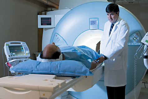
The photo shows a patient lying on a
sliding table outside of an MRI machine. The table will slide into the machine,
and the patient will lie quietly while pictures of the chest are taken.
You will hear loud humming, tapping, and buzzing
noises from the MRI machine. You may be able to use earplugs or listen to music
during the test.
Moving your body can cause the pictures to blur. The
technician will ask you to remain very still during the test. If you
can’t lie still, you may be given medicine to help you relax. The
technician also may ask you to hold your breath for 10 to 15 seconds at a time,
while he or she takes pictures of the structures in your chest.
What To Expect After Chest MRI
You usually can return to your normal routine right
after a chest MRI.
If you got medicine to help you relax during the
MRI, your doctor will tell you when you can return to your normal routine. The
medicine may make you tired, so you’ll need someone to drive you home.
If contrast dye was used during the test, you may
have a bruise where the needle was inserted. Also, if you’re
breast-feeding, you’ll need to bottle-feed your baby for a short time
after the test. The contrast dye can be passed to your baby through your breast
milk.
Ask your doctor how long you need to wait before you
breast-feed. You may want to prepare for the test by pumping and saving milk
for 24 to 48 hours in advance.
What Does a Chest MRI Show?
A chest MRI may show a tumor, problems in the blood
vessels (such as an
aneurysm
or blood clot), abnormal lymph nodes, and other chest conditions.
What Are the Risks of Chest MRI?
There are no risks from the magnetic fields or radio
waves used during a chest MRI.
Serious reactions to the contrast dye used for some
MRIs are very rare. However, side effects are possible and include the
following:
- Headache
- Nausea (feeling sick to your stomach)
- Dizziness
- Changes in taste
- Allergic reactions, such a hives and itchy eyes
Rarely, contrast dye is harmful to people who have
severe kidney disease.
Key Points
- Chest MRI is a safe, noninvasive test. It creates
detailed pictures of the structures in your chest, like your chest wall, heart,
and blood vessels. Radio waves, magnets, and a computer are used to make these
pictures.
- Chest MRI is used to look for tumors in the
chest; look at blood vessels, lymph nodes, and other structures in the chest;
and help explain results from other tests, such as
chest
x ray and chest CT scan.
- You may need a chest MRI if your doctor suspects
you have a chest condition, such as a tumor, a problem in the blood vessels
(such as an
aneurysm
or blood clot), abnormal lymph nodes, or other chest conditions.
- Before a chest MRI, your doctor or the MRI
technician will ask you questions about your health to make sure an MRI is safe
for you. You should not wear or bring metal or electronic objects into the MRI
room. The MRI can damage these items, and they can interfere with the MRI
machine.
- An MRI machine looks like a long, narrow tunnel.
New machines are shorter and wider and don’t completely surround you;
others are open on all sides. Tell your doctor if you’re afraid of tight
or closed spaces. He or she may give you medicine to help you relax or find you
a place that has an open MRI machine.
- Before the test, your doctor may inject a special
substance (called contrast dye) into a vein in your arm. This dye allows the
MRI to take more detailed pictures of the structures in your chest.
- If you’re breast-feeding, ask your doctor
how long you should wait after the test before you breast-feed. The contrast
dye can be passed through your breast milk. You may want to prepare for the
test by pumping and saving milk for 24 to 48 hours in advance. You can
bottle-feed your baby in the hours after the MRI.
- A chest MRI is painless. During the test, you lie
on your back on a sliding table as it passes through the MRI machine. The
machine takes pictures of your chest. Moving your body can cause the pictures
to blur. You will be asked to remain very still during the test.
- You usually can return to your normal routine
right after a chest MRI. If you got medicine to help you relax, your doctor
will tell you when you can return to your normal routine. The medicine may make
you tired, so you’ll need someone to drive you home.
- Chest MRI has few risks. Rarely, the contrast dye
used for some chest MRIs may cause an allergic reaction.
Links to Other Information About Chest MRI
Non-NHLBI Resources
|