|

Discovery
Safer Nano Cancer Detector

Nanoparticle test in mice could pave the way for human uses
April 30, 2009
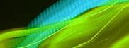
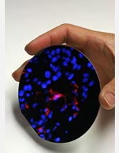
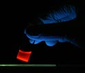
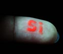
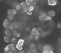
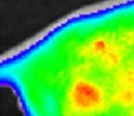
The first biodegradable fluorescent nanoparticle to safely image tumors and organs in live mice could be used for cancer detection and treatment in humans. Chemistry professor Michael Sailor and a team including National Science Foundation supported researchers at the University of California, San Diego, report developing the first nanoscale "quantum dot" particle that glows brightly enough to allow physicians to examine internal organs and lasts long enough to release cancer drugs before breaking down into harmless by-products. The research is another step towards mainstreaming the use of nanotechnology in medicine. Many researchers say using nanomaterials for medical reasons is the health field's next major frontier. The payoff, they say, could be lower drug toxicity, lower treatment costs, more efficient drug use, and better patient diagnosis. "There are a lot of nanomaterials that have an ability to do fluorescence imaging," says Sailor, referring to a useful property that potentially could help doctors further see organs, diagnose patients and perform surgeries. "But they're generally toxic and not appropriate for putting into people." The problem results from toxic organic or inorganic chemicals used to make the materials glow. For example, fluorescent semiconductor nanoparticles known as quantum dots can release potentially harmful heavy metals when they break down. A paramount issue in determining the efficacy of nanomaterials is the body's ability to harmlessly get rid of residual leftovers after the nanomaterial helps diagnose or treat a disease. So Sailor's team designed a new, non-toxic quantum dot nanoparticle made from silicon wafers, the same high-purity wafers that go into the manufacture of computer chips. Reseachers took the thin wafers and ran electric current through them drilling billions of pores. They then used ultrasound waves to break the wafer into bits as small as 100 nanometers. The resulting spongy silicon particles contained nano-scale features capable of displaying quantum confinement effects, or the so-called "quantum dots." The ones in the UCSD experiment glowed a reddish color when exposed to red, blue, or ultraviolet light. When the nanoparticles were tested in mice, researchers saw tumors glow for several hours, then dim as the particles degraded. The number of nanoparticles dropped noticeably in a week, and they were undetectable after four weeks. They performed a battery of toxicity assays and saw no evidence of toxicity. However, the researchers stopped short of concluding these new nanoparticles were completely harmless. "Very high doses of any substance can be harmful," says Sailor. "The important conclusion from this work is that the materials are nontoxic at the concentrations we need to use to see tumors." The fact that their quantum dots are made from silicon is key. "A major contributing factor is the fact that these materials degrade into silicic acid, a form of silicon that is commonly present in the human body and that is needed for proper bone and tissue growth," Sailor says. Examples where such materials should be useful include the early diagnosis and treatment of cancer. Nanoparticles that glow can reveal tumors too small to detect by other means. During surgery, they can allow the doctor to better find and remove all traces of a cancerous growth. In addition, they can enable targeted delivery of drugs and make it possible to use smaller, safer doses. Some cancer drugs such as doxorubicin, which is used in chemotherapy, can stick to the pore walls in the new biodegradable nanomaterial and slowly escape as the silicon dissolves. When doxorubicin is delivered to the whole body in doses high enough to be effective, it often has toxic side effects, and its incorporation in the new silicon nanoparticles may provide a more effective, less dangerous way to deliver this important drug. More needs to be done before this new material can undergo clinical trials in humans. Researchers need to further test its toxicity, how well it delivers drugs to diseased tissues, and how well it can be imaged in clinical settings. Graduate students Ji-Ho Park and Luo Gu in Sailor's lab; Sangeeta Bhatia, bioengineering professor at the Massachusetts Institute of Technology and graduate student Geoffrey von Malzahn in Bhatia's lab; and Erkki Ruoslahti, Tumor Microenvironment professor at the University of California, Santa Barbara assisted the research. Along with NSF, the National Cancer Institute helped fund the research.
| -- |
Bobbie
Mixon, National Science Foundation (703) 292-8070 bmixon@nsf.gov
|
| -- |
David
A. Brant, National Science Foundation (703) 292-4941 dbrant@nsf.gov
|
| -- |
Linda
S Sapochak, National Science Foundation (703) 292-4932 lsapocha@nsf.gov
|
Investigators
Michael Sailor
Sangeeta Bhatia
Erkki Ruoslahti
Related Institutions/Organizations
University of California-San Diego
Massachusetts Institute of Technology
University of California, Santa Barbara
Locations
California
Massachusetts
Related Programs
Biomaterials
Biomedical Engineering
Solid State and Materials Chemistry
Related Awards
#0452579 Chemistry of Nanostructured Porous Si
#9700202 Chemistry of Luminescent Porous Silicon
#9900034 Silicate Phosphors from Sol-Gel Prescursors
#0538512 CAREER: Microscale Structure/Function Studies Towards Development of Engineered Liver Tissue
#0806859 Materials World Network: "New Functionalized Hybrid Systems for Biosensing and drug Delivery"
Years Research Conducted
2008
Total Grants
$1,062,241
Related Agencies
National Cancer Institute
Related Websites
Sailor Research Group: http://sailorgroup.ucsd.edu/
Article in Nature Materials: http://www.nature.com/nmat/journal/v8/n4/full/nmat2398.html
|