|
National Institutes of Health |
|
|
|
Quick Links
NCRR and the 2009 Recovery Act
|
Laser Biomedical Research CenterON THIS PAGE: SEE ALSO:
OTHER OPTICAL AND LASER TECHNOLOGY CENTERS: Laser Biomedical Research Center
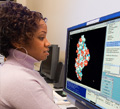
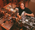
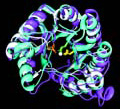
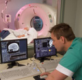
Research EmphasisThe Laser Biomedical Research Center develops methods of spectral diagnosis for in vivo analysis of diseased biological tissue; studies light propagation in turbid media by means of photon migration; develops spectroscopic techniques for imaging functional change in biological tissue; uses low-coherence interferometry to measure nanometer-scale length changes and dynamic processes in living cells and tissues; develops near-infrared Raman spectroscopy for accurate concentration measurements of blood analytes, diagnosis of breast cancer, and in vivo studies of atherosclerosis; and studies intrinsic noise in gene regulatory networks. The Laboratory of Neuro Imaging Resource (LONIR) develops novel strategies to investigate brain structure and function in their full multidimensional complexity. There is a rapidly growing need for brain models comprehensive enough to represent brain structure and function as they change across time in large populations, in different disease states, across imaging modalities, across age and gender, and even across species. International networks of collaborators are provided with a diverse array of tools to create, analyze, visualize, and interact with models of the brain. A major focus of these collaborations is to develop 4-dimensional brain models that track and analyze complex patterns of dynamically changing brain structure in development and disease, expanding investigations of brain structure-function relationships to four dimensions. Current ResearchCurrent research includes development of point contact and imaging instruments for use in a clinical setting. These systems are used to study reflectance, fluorescence, elastic light scattering, and Raman spectra from human tissues. Trimodal spectroscopy—a clinical technique that combines intrinsic fluorescence spectroscopy, diffuse reflectance spectroscopy, and light-scattering spectroscopy for spectral diagnosis—is being developed and tested in the esophagus, colon, bladder, cervix, and oral cavity. Near-infrared Raman spectroscopy is being developed for transcutaneous measurement of blood analyte concentrations. Raman histochemistry of breast cancer and atherosclerosis is being studied. Optical fiber probes for in vivo Raman spectroscopy are being developed and clinical studies in femoral and carotid arteries are underway. Low-coherence interferometry is being developed to measure nanometer-scale dynamic processes in biological systems. Instrumentation for ultrasensitive fluorescence microscopy to explore stochastic gene expression by a single gene is also being developed. Resource CapabilitiesInstruments and Experimental Set-UpsThe center houses state-of-the-art facilities for near-infrared, visible, and ultraviolet Raman spectroscopy, with micro-Raman capability and optical fiber probes for remote measurements; a ps/fs laser laboratory with a mode-locked Ti:sapphire laser with second, third, and fourth harmonic generation and an optical parametric amplifier to generate tunable radiation in the visible; a streak camera capable of 2 ps resolution; a fast-gated charge-coupled device (up to 100 MHz); instrumentation for fluorescence microspectroscopy; spectral endoscopes for fluorescence imaging of disease; and excitation-emission matrix spectrometers with multiwavelength excitation that collect an entire excitation-emission matrix in less than one second. Publications
|
| National Institutes of Health (NIH) Bethesda, Maryland 20892 |
Department of Health and Human Services |