|
National Institutes of Health |
|
|
|
Quick Links
NCRR and the 2009 Recovery Act
|
Resource for Magnetic Resonance and Optical ImagingON THIS PAGE: SEE ALSO:
IMAGING CENTERS:
Resource for Magnetic Resonance and Optical Imaging
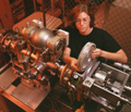
Research EmphasisThe focus of this resource is on developing instrumentation, methodologies, and data analysis techniques for the quantitative assessment of functional, structural, and metabolic parameters in humans with the use of multinuclear magnetic resonance, novel spectral, perfusion, functional, and optical imaging techniques. Current ResearchIn conjunction with our collaborators, the resource is pursuing the following four broad areas of core research. The first core deals with the development of novel magnetic resonance (MR) techniques for studying the structural, biochemical, and metabolic aspects of cartilage, brain, and tumors, with direct application to arthritis, stroke, Alzheimer's disease, and cancer. This core also develops novel image reconstruction strategies to quantify high temporal contrast agent dynamics in breast and other tissues. In the second core, research is being undertaken to improve quantitative perfusion imaging at high fields and in pediatric patients as well as methods for concurrent imaging of blood flow and glucose metabolism. It also develops strategies for correlation of functional magnetic resonance imaging (fMRI) with optical imaging. The third core's subprojects deal with MR of hyperpolarized gases and development of a comprehensive approach for the study of pulmonary and sinus diseases. This core also develops strategies for improving the efficiency of hyperpolarization of 129Xe. The fourth and final core focuses on combining optical and MRI techniques, development of methods of two-photon optical metabolic imaging and image reconstruction strategies in diffusion tomography for the study of neurophysiology, and breast cancer. The facility's core sections provide research and computing resources for numerous user, collaborative, and training projects. Resource Capabilities1.5 T Siemens Sonata, a 3 T Siemens Trio, and a 2 T, 1-meter bore, magnet system with a versatile spectrometer for multinuclear spectroscopy and imaging with in-magnet exercise capability; 4.7 T (30 cm diameter) and 9.4 T (10 cm diameter) vertical magnets for animal imaging; specialized radiofrequency probes for various nuclei; local magnetic field gradient sets; Workstations for data analysis; electronic test equipment; physiological monitoring equipment; in-magnet exercise apparatus; and bioelectric amplifiers and recorders. Metabolic spectroscopy including (but not limited to) 1H, 13C, 15N, 23Na, 7Li, 19F, 31P, and 17O, equipped for physiologic synchronization (gating) and decoupling, magnetization transfer experiments, two-dimensional, multiple quantum spectroscopy, and in-magnet exercise. Software and hardware support for all nuclear magnetic resonance experiments, including sodium and phosphorus imaging, chemical shift imaging, Hadamard spectroscopic imaging, 3He, and 126Xe imaging, and high-resolution proton imaging and flow. Facilities and expertise for generating hyperpolarized helium and xenon gases, specialized coil design, and construction are available, as are body-positioning devices for specific experiments (e.g., in-magnet exercise and heart, brain, and liver studies). Expertise and infrastructure for state-of-the-art functional perfusion imaging and integrated optical and MRI experiments. InstrumentsWhole-body MRI Scanners: 1.5 T Siemens Sonata whole-body clinical scanner, 2 T custom-built whole-body system, 3 T Siemens Trio whole-body clinical scanner with multinuclear capability, 4.7 T/50 cm Varian small-bore MRI system, 7.1 T Bruker AMX-300 Spectrometer, 9.4 T/8.9 cm Varian small-bore MRI system, and 11.8 T Bruker AMX-500 Spectrometer. SoftwareMATPULSE Extractor BreastPro TIMESERIES CYLINDER O2Analysis-pro Pulse sequences HardwareHardware designs for T/R switches and radiofrequency coils are also available. For more information, please contact Ari Borthakur at ari@mail.mmrrcc.upenn.edu. FacilitiesFacilities exist for building radiofrequency and gradient coils based on the demand of projects. The MMRRCC houses an electronics lab and has access to dedicated coil building facilities in both the Department of Radiology and an adjacent NMR facility. Personnel are available for coil building and consulting in coil design. The MMRRCC also maintains a chemistry laboratory, required for biological MR research and a mechanical shop for fabrication of custom, non-magnetic components for various specialized needs. The MMRRCC also has facilities for producing hyperpolarized noble gases using various lasers and other equipment. Associated centers and laboratories include the Center for Advanced Magnetic Resonance Imaging and Spectroscopy, the Center for Functional Neuroimaging, the Laboratory for Multinuclear Magnetic Resonance Imaging, and the Laboratory for Molecular Imaging. Training OpportunitiesTrainees and researchers come from a variety of backgrounds, including medicine, bioengineering, biophysics, chemistry, physics, engineering, and mathematics. The resource center excels at educating future scientists on the fundamentals of magnetic resonance theory and applications. Both new and established researchers are aided in learning about new developments in the field and to hone their skills for applications within their own research programs. Investigators may be trained on an individual basis by resource staff or through group mechanisms such as seminars, courses, and workshops. The resource facility houses more than 40 desks in a wall-less, cubicle-free environment that promotes open communication and discussion among the Principal Investigator, faculty members, predoctoral and postdoctoral fellows, and visiting and senior researchers. Publications
|
| National Institutes of Health (NIH) Bethesda, Maryland 20892 |
Department of Health and Human Services |