Add to My Images
Low-Resolution
Image
193 × 113 pixels
2.681 × 1.569 inches (72 dpi)
9.16 KB
view
download
Medium-Resolution
Image
402 × 235 pixels
2.680 × 1.567 inches (150 dpi)
34.93 KB
view
download
High-Resolution
Image
803 × 469 pixels
2.677 × 1.563 inches (300 dpi)
441.70 KB
view
download
|
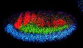
Neural development
ID Number
2327
Description
Using techniques that took 4 years to design, a team of developmental biologists has shown that certain proteins can direct the subdivision of fruit fly and chicken nervous system tissue into the regions depicted here in blue, green, and red. Molecules called bone morphogenetic proteins (BMPs) helped form this fruit fly embryo. While scientists knew that BMPs play a major role earlier in embryonic development, they didn't know how the proteins help organize nervous tissue. The findings suggest that BMPs are part of an evolutionarily conserved mechanism for organizing the nervous system.
The National Institute of Neurological Disorders and Stroke also supported this work.
Featured in the October 17, 2006, issue of Biomedical Beat.
|
|
Type
Photograph
|
Source
Mieko Mizutani and Ethan Bier, University of California, San Diego, and Henk Roelink, University of Washington
|


