Types of Congenital Heart Defects
Congenital heart defects change the normal flow of
blood through the heart because some part of the heart didn’t develop
properly before birth.
There are many types of congenital heart defects.
They include simple ones such as a hole in the interior walls of the heart that
allows blood from the left and right sides of the heart to mix, or a narrowed
valve that blocks the flow of blood to the lungs or other parts of the
body.
Other defects are more complex. These include
combinations of simple defects, problems with where the blood vessels leading
to and from the heart are located, and more serious abnormalities in how the
heart develops.
Examples of Simple Congenital Heart Defects
Holes in the Heart (Septal Defects)
The septum is the wall that separates the chambers
on the left side of the heart from those on the right. It prevents mixing of
blood between the two sides of the heart. Sometimes, a baby is born with a hole
in the septum. When that occurs, blood can mix between the two sides of the
heart.
Atrial
septal defect (ASD). An ASD is a hole in the part of the septum
that separates the atria—the upper chambers of the heart. This heart
defect allows oxygen-rich blood from the left atrium to flow into the right
atrium instead of flowing to the left ventricle as it should. Many children who
have ASDs have few, if any, symptoms.
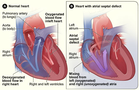
Normal Heart and Heart With Atrial
Septal Defect
Figure A shows the normal structure
and blood flow in the interior of the heart. Figure B shows a heart with an
atrial septal defect, which allows oxygen-rich blood from the left atrium to
mix with oxygen-poor blood from the right atrium.
An ASD can be small or large. Small ASDs allow only
a little blood to leak from one atrium to the other. Very small ASDs don’t
affect the way the heart works and therefore don’t need any special
treatment. Many small ASDs close on their own as the heart grows during
childhood. Medium to large ASDs allow more blood to leak from one atrium to the
other, and they’re less likely to close on their own.
Half of all ASDs close on their own or are so small
that no treatment is needed. Medium to large ASDs that need treatment can
usually be repaired using a catheter procedure. (See
“How Are Congenital Heart Defects
Treated?”)
Ventricular septal defect (VSD). A
VSD is a hole in the part of the septum that separates the ventricles—the
lower chambers of the heart. The hole allows oxygen rich blood to flow from the
left ventricle into the right ventricle instead of flowing into the aorta and
out to the body as it should.
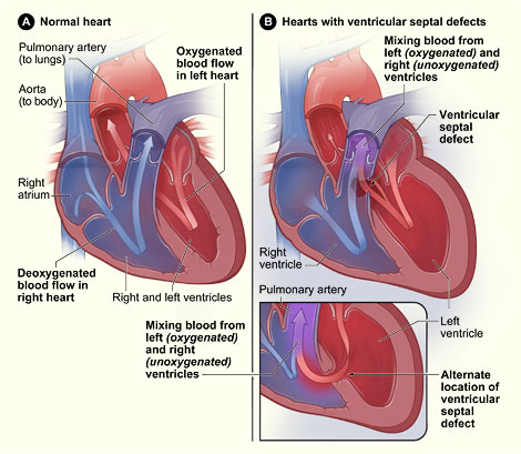
Normal Heart and Heart With
Ventricular Septal Defect
Figure A shows the normal structure
and blood flow in the interior of the heart. Figure B shows two common
locations for a ventricular septal defect. The defect allows oxygen-rich blood
from the left ventricle to mix with oxygen-poor blood in the right
ventricle.
A VSD can be small or large. A small VSD
doesn’t cause problems and may often close on its own. Large VSDs cause
the left side of the heart to work too hard and increase blood pressure in the
right side of the heart and the lungs because of the extra blood flow. The
increased work of the heart can cause
heart
failure and poor growth. If the hole isn’t closed, the high blood
pressure in the lungs can cause the delicate arteries in the lungs to scar, a
condition called
pulmonary
arterial hypertension. Open-heart surgery is used to repair VSDs.
Narrowed Valves
Simple congenital heart defects also can involve the
heart’s valves, which control the flow of blood from the atria to the
ventricles and from the ventricles into the two large arteries connected to the
heart (the aorta and the pulmonary artery). Valves can have the following types
of defects:
- Stenosis. This is when the valve doesn’t
open completely, and the heart has to work harder to pump the blood through the
valve.
- Atresia. This is when the valve doesn’t form
correctly, so there is no opening for blood to pass through.
- Regurgitation (re-GUR-ji-TA-shun). This is when
the valve doesn’t close completely, so blood leaks back through the valve.
The most common valve defect is called pulmonary
valve stenosis, which is a narrowing of the pulmonary valve. This valve allows
blood to flow from the right ventricle into the pulmonary arteries and out to
the lungs to pick up oxygen.
Pulmonary valve stenosis can range from mild to
severe. Most children with this defect have no signs or symptoms other than a
heart murmur. Treatment isn’t needed if the stenosis
is mild.
In a baby with severe pulmonary valve stenosis, the
right ventricle can get very overworked trying to pump blood to the pulmonary
arteries. Oxygen-poor blood can back up from the right side of the heart into
the left side, causing cyanosis. Cyanosis is a bluish tint to the skin, lips,
and fingernails. It occurs because the oxygen level in the blood leaving the
heart is below normal.
Older children with severe pulmonary valve stenosis
may have symptoms such as fatigue (tiredness) when exercising. Severe pulmonary
valve stenosis is treated with a catheter procedure.
Example of a Complex Congenital Heart Defect
Complex congenital heart defects need to be repaired
with surgery. Because of advances in diagnosis and treatment, doctors can now
successfully repair even very complex congenital heart defects.
The most common complex heart defect is
tetralogy of Fallot (teh-TRALL-o-gee of fall-O), a
combination of four defects:
- Pulmonary valve stenosis.
- A large VSD.
- An overriding aorta. The aorta sits above both
the left and right ventricles over the VSD, rather than just over the left
ventricle. As a result, oxygen poor blood from the right ventricle can flow
directly into the aorta instead of into the pulmonary artery to the lungs.
- Right ventricular hypertrophy. The muscle of the
right ventricle is thicker than usual because of having to work harder than
normal.
These defects prevent enough blood from flowing to
the lungs to get oxygen, while oxygen-poor blood flows directly out to the
body.
Normal Heart and Heart With
Tetralogy of Fallot
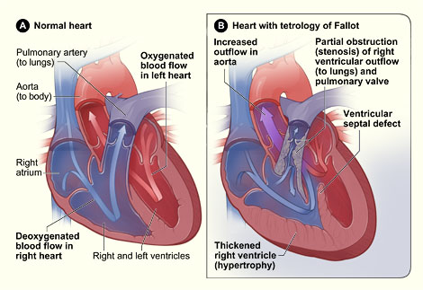
Figure A shows the normal structure
and blood flow in the interior of the heart. Figure B shows a heart with the
four defects of tetralogy of Fallot.
Babies and children with tetralogy of Fallot have
episodes of cyanosis, which can sometimes be severe. In the past, when this
condition wasn’t treated in infancy, older children would get very tired
during exercise and could have fainting spells. Tetralogy of Fallot is now
repaired in infancy to prevent these types of symptoms.
Tetralogy of Fallot must be repaired with open heart
surgery, either soon after birth or later in infancy, depending on how severely
the pulmonary artery is narrowed. Children who have had this heart defect
repaired need lifelong medical care from a specialist to make sure they stay as
healthy as possible.
|