What Is Carotid Ultrasound?
Carotid (ka-ROT-id) ultrasound is a painless and
harmless test that uses high-frequency sound waves to create images of the
insides of the two large arteries in your neck. These arteries, called carotid
arteries, supply your brain with blood. You have one carotid artery on each
side of your neck.
Carotid ultrasound shows whether a material called
plaque (plak) has narrowed your carotid arteries. Plaque is made up of fat,
cholesterol, calcium, and other substances found in the blood. It builds up on
the insides of your arteries as you age.
Too much plaque in a carotid artery can cause a
stroke. The plaque can slow down or block the flow of blood
through the artery, allowing a blood clot to form. A piece of the blood clot
can break off and get stuck in the artery, blocking blood flow to the brain.
This is what causes a stroke.
A standard carotid ultrasound shows the structure of
your carotid artery. Your carotid ultrasound test may include a Doppler
ultrasound. Doppler ultrasound is a special ultrasound that shows the movement
of blood through your blood vessels. Your doctor often will need results from
both types of ultrasound to fully assess if there is a problem with blood flow
through your carotid arteries.
Other Names for Carotid Ultrasound
- Doppler ultrasound
- Carotid duplex ultrasound
Who Needs Carotid Ultrasound?
Carotid ultrasound checks for plaque buildup in the
carotid arteries. This buildup can narrow or block your carotid arteries. You
may need a carotid ultrasound if you:
- Had a
stroke or ministroke recently.
- Have an abnormal sound in your carotid artery
called a carotid bruit (broo-E). Your doctor can hear a carotid bruit with the
help of a stethoscope put on your neck over the carotid artery. A bruit can
mean that there’s a partial blockage in your carotid artery that could
lead to a stroke.
Your doctor also may order a carotid ultrasound if
he or she suspects you may have:
- Blood clots that can slow blood flow in your
carotid artery
- A split between the layers of your carotid artery
wall that weakens the wall or reduces the blood flow to your brain
A carotid ultrasound also may be done to see whether
carotid artery surgery has restored normal blood flow. If you had a procedure
called carotid
stenting,
your doctor may order a carotid ultrasound afterward to check the position of
the stent put in your carotid artery. (The stent, a small mesh tube, helps
prevent the artery from becoming narrowed or blocked again.)
Sometimes carotid ultrasound is used as a preventive
screening test in people who have medical conditions that increase their risk
of stroke, including
high
blood pressure and diabetes. People with these conditions may benefit from
having their carotid arteries checked regularly even if they show no signs of
plaque buildup.
What To Expect Before Carotid Ultrasound
Carotid ultrasound is a painless test, and typically
there is little to do in advance. Your doctor will tell you how to prepare for
your carotid ultrasound.
What To Expect During Carotid Ultrasound
Carotid ultrasound is usually done in a
doctor’s office or hospital. The test is painless and usually doesn’t
take more than 30 minutes.
The ultrasound machine includes a computer, a video
screen, and a transducer, which is a hand-held device that sends and receives
ultrasound waves into and from the body.
You will lie down on your back on an exam table for
the test. Your technician or doctor will put a gel on your neck where your
carotid arteries are located. This gel helps the ultrasound waves reach the
arteries better. Your technician or doctor will put the transducer against
different spots on your neck and move it back and forth.
The transducer gives off ultrasound waves and
detects their echoes after they bounce off the artery walls and blood cells.
Ultrasound waves can’t be heard by the human ear.
A computer uses the echoes of the ultrasound waves
bouncing off the carotid arteries to create and record images of the insides of
the arteries (usually in black and white) and your blood flowing through them
(usually in color; this is the Doppler ultrasound). A video screen displays
these live images for your doctor to review.
Carotid Ultrasound
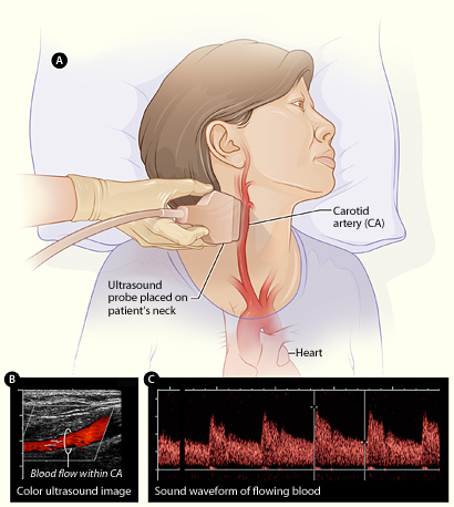
Figure A shows how the ultrasound
probe is placed over the carotid artery. Figure B is a color ultrasound image
showing blood flow (the red color in the image) in the carotid artery. Figure C
is a waveform image showing the sound of flowing blood in the carotid
artery.
What To Expect After Carotid Ultrasound
Usually there is nothing special you have to do
after a carotid ultrasound, and you should be able to return to normal
activities immediately.
Often your doctor will be able to tell you the
results of the carotid ultrasound when it occurs or soon afterward.
What Does a Carotid Ultrasound Show?
A carotid ultrasound can show whether buildup of a
fatty material called plaque has narrowed one or both of your carotid arteries
and reduced blood flow to your brain.

The illustration shows a normal
artery with normal blood flow (figure A) and an artery containing plaque
buildup (figure B).
If your carotid arteries are narrowed by plaque, you
may be at risk for having a
stroke. That risk depends on how much of your artery is
blocked and how much blood flow is restricted. To reduce your risk for stroke,
your doctor may recommend medical or surgical treatments to reduce or remove
the plaque buildup in your carotid arteries.
What Are the Risks of Carotid Ultrasound?
There are no risks linked to having a carotid
ultrasound, because the test uses harmless sound waves. These are the same type
of sound waves that doctors use to record pictures of fetuses in pregnant
women.
Key Points
- Carotid ultrasound is a test that uses
high-frequency sound waves to create images of the insides of the two large
arteries in your neck. These arteries, called carotid arteries, supply your
brain with blood.
- A carotid ultrasound can show whether buildup of
a fatty material called plaque has narrowed one or both of your carotid
arteries and reduced blood flow to your brain.
- If your carotid arteries are narrowed by plaque,
you may be at risk for having a
stroke, depending on how much of your artery is blocked and
how much blood flow is restricted.
- You may need a carotid ultrasound if you had a
stroke or ministroke recently or are at high risk for having a stroke.
- Carotid ultrasound is a painless test done in a
doctor’s office or hospital. It usually doesn’t take more than 30
minutes and requires no preparation or followup.
- There are no risks linked to having a carotid
ultrasound, because the test uses harmless sound waves.
Links to Other Information About Carotid
Ultrasound
NHLBI Resources
- Stents
(Diseases and Conditions Index)
Non-NHLBI Resources
Clinical Trials
|