February 22, 2005
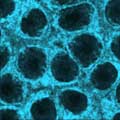
Cool Image: Movements of Myosin
Inside the fertilized egg cell of a fruit fly, we see a type of myosin, related to the protein that helps our muscles contract, made to glow by attaching a fluorescent protein. At the start of the movie, the myosin proteins are distributed relatively evenly near the surface of the embryo. The proteins temporarily vanish each time the cell's nuclei--at this point buried deep in the cytoplasm--divide. When the multiplying nuclei move to the surface, they shift the myosin, producing darkened holes. The glowing myosin proteins then gather, contract, and start separating the nuclei into their own compartments. Courtesy of Victoria Foe, research professor at the Center for Cell Dynamics, University of Washington's Friday Harbor Laboratories.
Time-lapse movie (20.9MB MOV)
Foe home page
Center for Cell Dynamics home page
More from this issue
Biomedical Beat is produced by the Office of Communications and Public Liaison of the National Institute of General Medical Sciences. For more information about the Institute, visit http://www.nigms.nih.gov. Some of the research briefs in this digest were generated from university or national laboratory news releases. For more information about Biomedical Beat, please contact the editor, Emily Carlson, at carlsone@nigms.nih.gov or 301-594-1515. To talk to someone at NIGMS about this research, call 301-496-7301. The material in this newsletter is not copyrighted and we encourage its use or reprinting. |