


|
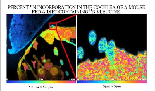
Multi-Isotope Imaging Mass Spectrometry (MIMS) used to image a mouse cochlea This image shows the percentage of the stable (non-radioactive) nitrogen isotope, 15N, incorporated into a mouse cochlea. The cochlea is a structure within the mouse’s inner ear. The mouse was fed a diet containing food with a protein precursor amino acid, l-leucine, which was enriched with 15N. Using the method of Multi-Isotope Imaging Mass Spectrometry (MIMS) developed at the Brigham and Women’s Hospital National Resource for Imaging Mass Spectrometry, scientists imaged and measured the localization of new proteins in the cochlea. Called “an imaging revolution” (Weitzman, 2006, J. Biol. 5:17), this highly sensitive technique allows for simultaneous measurement of molecular accumulation and distribution within cells. Image courtesy of Dr. Claude Lechene, Brigham and Women’s Hospital. EB 001974
|

 |
 |
Department of Health and Human Services |
 |
National Institutes of Health |
 |
