|
Magnetic Resonance Imaging (MRI) uses radio waves in the presence of a strong magnetic field that surrounds the opening of the MRI machine where the patient lies to get tissues to emit radio waves of their own.
Different tissues (including tumors) emit a more or less intense signal based on their chemical makeup, so a picture of the body organs can be displayed on a computer screen. Much like CT scans, MRI can produce three-dimensional images of sections of the body, but MRI is sometimes more sensitive than CT scans for distinguishing soft tissues.
 |
|
|
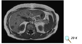
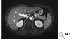
(Top) MRI scan without contrast showing possible tumor in the liver. (Bottom) MRI scan of the same patient using contrast. Images courtesy of Dr.
Peter Choyke, Clinical Center, NIH. |
< Previous | Next Section > Main |