


Home > Health & Education > eAdvances
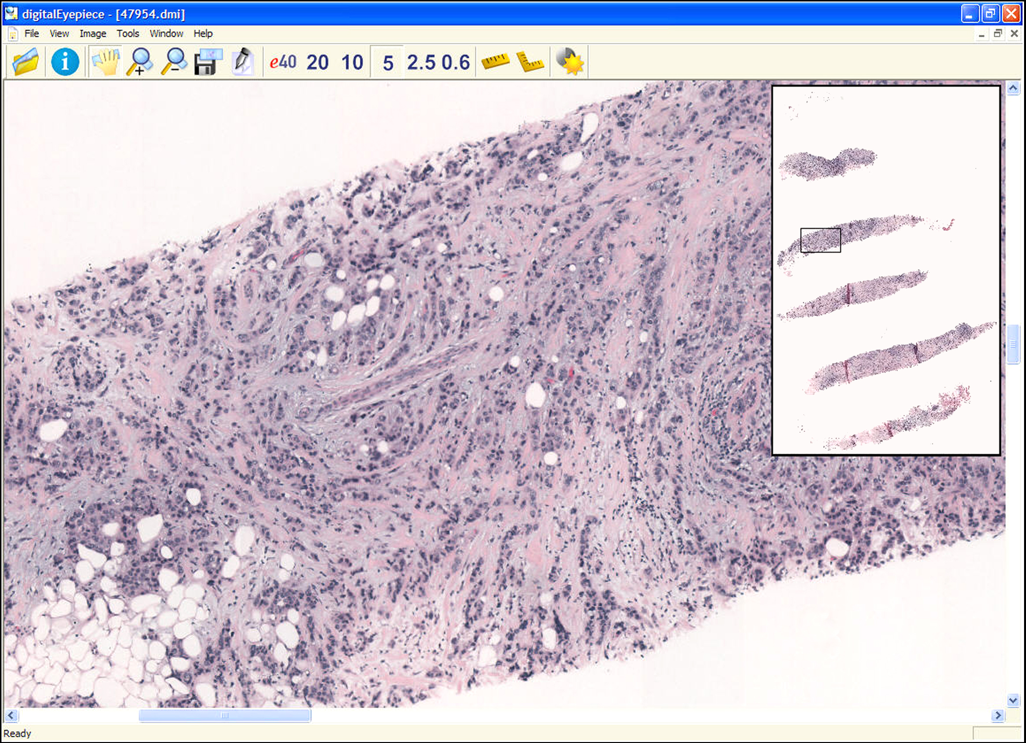
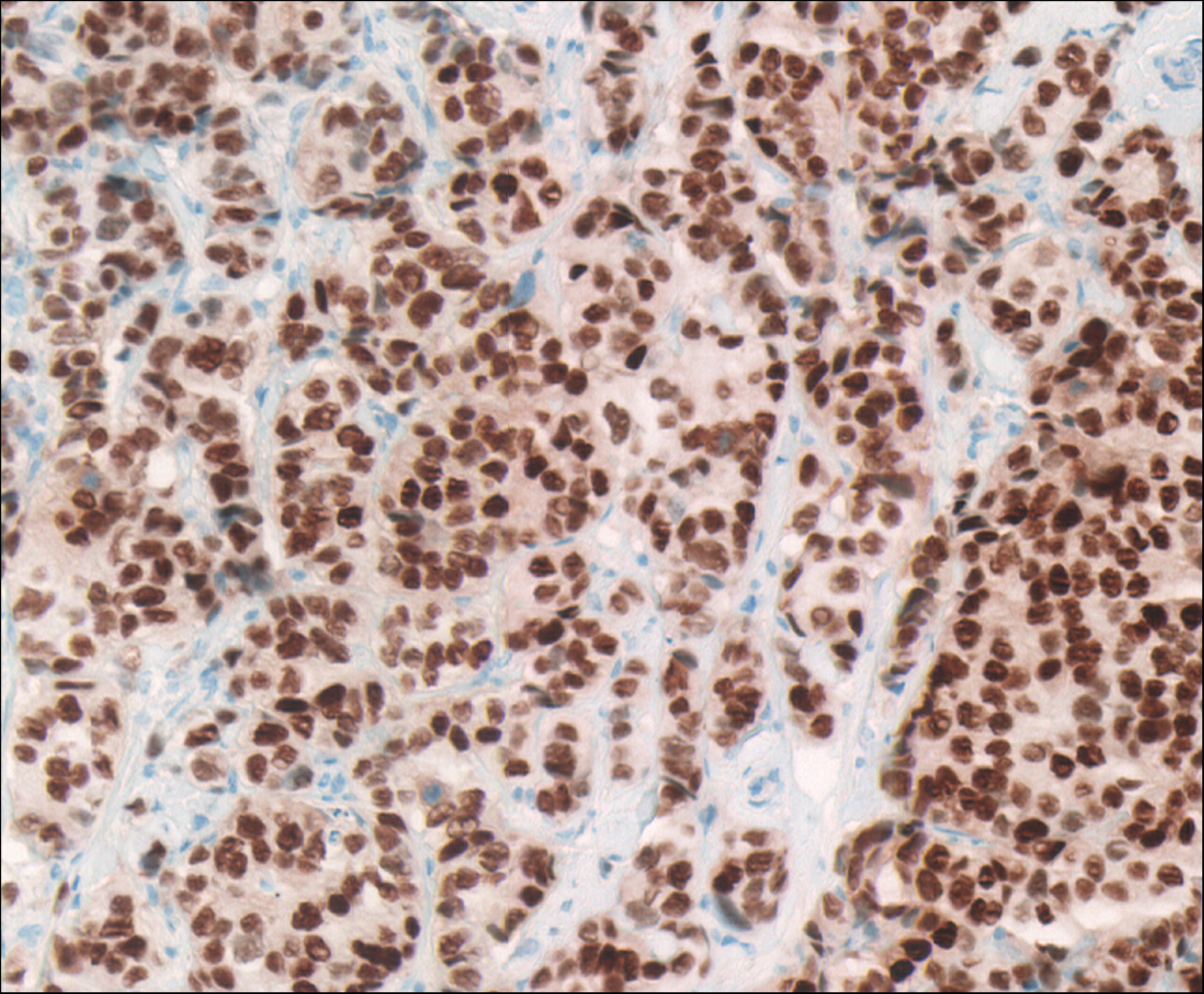
Faster Biopsy Results with New Microscope Array: August 30, 2006Even the most confident female patient gets rattled waiting a week or two for breast biopsy results. The tissue must be processed, examined by a pathologist, and a report sent to a surgeon or mammographer who shares the results with the patient. If the pathologist diagnoses a breast malignancy, another week or two may go by if the patient seeks a second opinion. A new approach developed at the University of Arizona College of Medicine allows patients to have their biopsy processed, interpreted, diagnosed and a second opinion rendered in one day. By combining rapid tissue processing with telepathology and teleoncology, cancer diagnosis times have dropped to a matter of hours, according to Ronald S. Weinstein, M.D., head of the University of Arizona’s Department of Pathology. At the heart of the system is a virtual pathology slide scanner developed by a team of engineers at Tucson-based DMetrix Inc. The scanner features a novel microscope that includes an array of 80 tiny microscopes which simultaneously scan across a whole piece of tissue. "What comes out of our system is a whole slide digital image in one pass," says Artur Olszak, director of engineering at DMetrix. "The process takes less than a minute." The Conventional WayConventional virtual slide scanners use a single optical-axis light microscope to scan a section of tissue mounted on a glass slide. But pathologists’ need for very high-resolution images means that the tissue section is digitally imaged one small area at a time. Single-optical axis microscope-based digital imaging systems scan many fields individually and then stitch them together to create a composite image. The entire process for scanning just one glass histopathology slide takes between five and 60 minutes. "There is a fundamental trade-off between field-of-view and resolution present in conventional optics," Olszak says. "You either can view a large field-of-view with low resolution or vice versa: high resolution with a smaller field-of-view." Building An ArrayTo get around this problem, DMetrix engineers created a new type of optical instrument that is based on the concept of a lenslet array ensemble. This consists of three-tiered, stacked arrays of lenses with each lens just 1.5 mm in diameter, Olszak says. Each lenslet array is the size of a U.S. quarter. Eight rows of 10 lenses are staggered relative to each other to fit them into a very small area. A tiny camera is attached to the top of the lenslet array ensemble. The entire lenslet array ensemble including the camera/sensor is the size of a stack of five U.S. quarters. The ensemble hovers over, and moves along, the long axis of the glass slide. "This is a new class of optical systems that produces very good image quality," Olszak says. Automating the scanning process helped reduce time to diagnosis as well. Once a laboratory technician loads a set of slides into a cassette, the machine can do the rest: "The system can determine where the tissue is, focuses automatically, deposits the files in the folder, and then you can see them online," Olszak says. "The scanner is fully independent - it's called walk-away scanning." The resulting images can be shared among pathologists working in the same hospital or with off-site pathologists at distant locations. Efficient digital pathology will also be invaluable in the classroom, Olszak says, so that instructors can make sure all students see features in the same microscopic tissue samples. DMetrix virtual slides have been incorporated into the curriculum at the University of Arizona College of Medicine and are popular among medical students. Helping PatientsDoctors in the University of Arizona's University Medical Center pathology practice have been using the DMetrix system for more than a year to evaluate breast biopsies as part of a research project. "We've done dozens of DMetrix cases and women are delighted with this same day biopsy service," says Weinstein who also is director of the state-wide Arizona Telemedicine Program and a co-founder of DMetrix, Inc. Through UltraClinics®, a telemedicine service provider company also founded by Weinstein, the rapid scanning technology will benefit patients in remote areas since their virtual slides can be posted on a server and accessed on a secure Internet site. In addition to breast biopsies, UltraClinics® also provides rapid prostate cancer diagnoses. Future PossibilitiesIt’s possible that the miniaturized microscope array technology could be used in fields outside of pathology. “We talk only about medicine, but the same principle of fast acquisition and high resolution can be used on a semiconductor wafer, for example,” Olszak says. “There are a lot of different areas that we haven't even touched." In recognition of the new technology, DMetrix’s first product, the DX-40Array Microscope, won a 2005 R&D 100 Award from R&D Magazine and was runner-up in The Wall Street Journal’s 2005 Global Technology Innovation Awards. The DMetrix DX-40 Array Microscope was developed with support from a Small Business Innovative Research (SBIR) grant from the National Institute of Biomedical Imaging and Bioengineering (NIBIB). References: Olszak, AG, and MR Descour, Microscopy in multiples, oemagazine, 5: 16-18, 2005.Weinstein, RS, Innovations in medical imaging and virtual microscopy, Human Pathology, 36: 317-319, 2005. Weinstein RS, et al., Reinvention of light microscopy. Array microscopy and ultrarapidly scanned virtual slides for diagnostic pathology and medical education. In: Virtual microscopy and Virtual Slides in Teaching, Diagnosis and Research, J Gu and RW Ogilvie, eds., CRC Francis and Taylor Press, 9-34, 2005. Kayser K, et al., Telepathology and Telemedicine: Communication, Electronic Education and Publication in e-Health. VSV Interdisciplinary Medical Publishing, Berlin, 23-29, 2005. Weinstein RS, et al., An array microscope for ultrarapid virtual slide processing and telepathology, Human Pathology, 35:1303-1314, 2004. UltraClinics® is a registered trademark of UltraClinics®, Inc.
|


 |
 |
Department of Health and Human Services |
 |
National Institutes of Health |
 |
