Home > Health & Education > eAdvances
E-mail this page 

Novel PET/MRI Scanner Expands Cancer and Drug Studies: May 28, 2008
As the medical community begins to embrace the concept of personalized medicine – a tailoring of therapeutic treatments to an individual – new tools are needed to monitor how the body responds to both disease and treatment.
One such tool, positron emission tomography combined with computed tomography (PET/CT), is gaining wider acceptance in clinical settings because of its ability to pinpoint and assess cancer, heart disease, and brain disorders. PET/CT images provide clinicians with information on structure (from CT) as well as tissue function (from PET). PET/CT, however, has several drawbacks: repeat scans increase the amount of ionizing radiation to which patients are exposed, CT does not image soft tissue as well as magnetic resonance imaging (MRI), and CT does not have the same degree of flexibility in providing functional and physiologic information as MRI.
A research team led by Simon Cherry, Professor of Biomedical Engineering at the University of California, Davis, has developed a new imaging system that answers PET/CT’s limitations. They’ve paired PET with nonionizing MRI in a single system that simultaneously acquires MRI and PET data. Combining the imaging approaches, however, was no simple feat since the two systems can readily interfere with each other and produce poor-quality images.
|
|
|
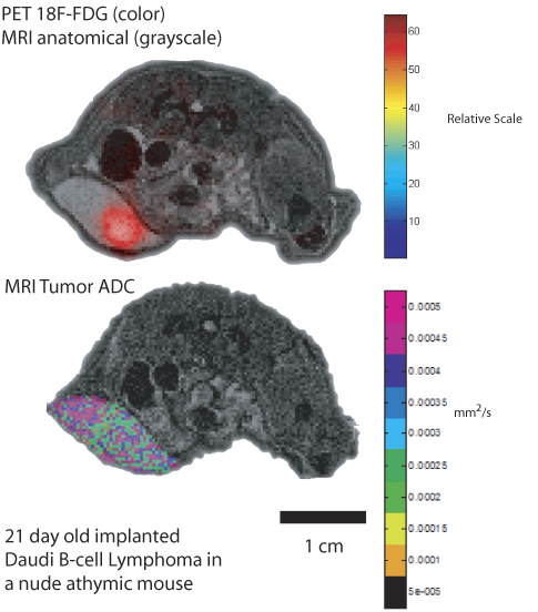
A combined PET/MRI image shows a lymphoma tumor in a mouse's groin area. The top image shows localization (red) of an imaging agent (18F-FDG) in the tumor on top of a simultaneously recorded MR image showing bowels (dark ovals) and spinal cord (center top bright spot). The bottom MR image (recorded minutes later) shows a quantitative map of diffusive water movement within the tumor. Image courtesy of Thomas Ng and Daniel Procissi, California Institute of Technology, Pasadena.
|
|
The Combination Challenge
PET tracks the movement of specially designed molecules called radiotracers through the body, giving information on the concentrations of these molecules, but does not readily pinpoint the anatomic location of the signal. MRI provides high-resolution images of soft tissue using a strong magnetic field. Researchers use both approaches in clinical and basic research.
Problems arise when investigators try to correlate data from PET and MRI. Images obtained on separate systems must be aligned using software that looks for identical landmarks on the two images, a challenge when it comes to organs in the thorax and abdomen that can easily be displaced or change shape when a patient is moved from one scanner to another. Separate systems also mean that data are gathered at different points in time. Drug uptake or distribution of an imaging agent often occurs in a matter of seconds or minutes, leaving little time to switch from a PET to an MRI scanner.
With these issues in mind, Cherry and his team of engineers from the University of California, Davis, California Institute of Technology, and University of Tübingen, designed and built a PET insert that slides into an MRI scanner. Components were chosen to minimize electronic and magnetic interference between the two systems.
“The combined PET/MRI scanner provides highly complementary information that allows you to study different aspects of a biological system aligned in both space and time,” explains Cherry. “Because biological systems are complex, dynamic systems, this provides powerful opportunities to ask questions about the validation of new therapeutics, diagnostics, and biomarkers that are very hard to answer using separate imaging systems.”
Nonclinical Imaging Tool
Cherry anticipates that the PET/MRI system will become a key tool in nonclinical animal research. In such drug studies, the imager would allow researchers to track the effectiveness of a drug over time in the same animal, reducing the number of animals used in an experiment. “We’ll be able to achieve the same level of statistical significance with far fewer animals,” he says.
Studies by Cherry’s team on mice with implanted tumors have demonstrated PET/MRI’s ability to differentiate between malignant tissue, dead tissue, and swelling with a tumor. The researchers have also performed whole-body imaging with the system. This kind of imaging allows assessment of structural and physiological changes across organ systems. In studies of cancer metastasis (the migration of tumor cells to areas distant from the original cancer site), PET can locate the breakaway cells and MRI can provide structural or functional information on exactly where the metastasis is located.
“The goal in animals is better understanding of disease progression and of how treatments interact with an animal model,” says Cherry. “The earlier we can detect the effects of a therapeutic intervention [in animals], the better. This will allow us to accelerate and inform the choice as to which therapeutic strategy should be moved to human trials and, eventually, stratify who should go on which drugs. This will help build the foundations for personalized medicine.”
Potential Clinical Tool
While PET/MRI images offer keen views of tissues and organs, unique imaging dyes or contrast agents clarify structures with unprecedented precision. Those agents may hold the key to the hybrid system’s wide acceptance as a clinical tool. To work out this issue, Cherry tapped UC Davis colleague Angelique Louie, an Associate Professor in the Department of Biomedical Engineering, who specializes in designing novel imaging agents. She has created a number of novel agents that carry both paramagnetic ions for MRI and positron-emitting ions for PET. These compounds will allow the researchers to take full advantage of MRI’s high spatial resolution to view tissue structures and PET’s high sensitivity to view concentrations of cells.
One application Louie is studying involves imaging plaques or fatty deposits in blood vessels. Research has shown that plaques are more likely to rupture if they have high concentrations of macrophages (cells that engulf and destroy bacteria and other foreign matter) and specific patterns of macrophage distribution. A plaque rupture can create life-threatening complications such as heart attack or stroke.
“It is now understood that it is not the size of the plaque but the likelihood of rupture that is the cause of major adverse health effects,” explains Louie. To image plaques Louie created agents that target macrophages. When injected into the body, the contrast agents accumulate in these cells. “We envision that PET would be used for screening to identify the most vulnerable lesions, and then the high resolution of MRI would be used to assess the macrophage distribution and density in these plaques.”
Although Cherry is cautiously optimistic about PET/MRI’s potential for widespread clinical use, Louie anticipates a swift transition. “PET/CT is the fastest growing area of sales in imaging instruments and has been increasingly adopted in the clinic,” says Louie. With human-scale PET/MRI instruments now in development by manufacturers of clinical imaging systems, “PET/MRI looks poised to follow a trajectory similar to PET/CT.”
This work is supported in part by the National Institute of Biomedical Imaging and Bioengineering.
References
Catana C, Procissi D, Wu Y, Judenhofer MS, Qi J, Pichler BJ, Jacobs RE, Cherry SR. Simultaneous in vivo positron emission tomography and magnetic resonance imaging. Proc Natl Acad Sci. 2008 Mar 11;105(10):3705-10.
Cherry SR, Louie AY, Jacobs RE. The integration of positron emission tomography with magnetic resonance imaging. Proc IEEE. 2008 Mar;96(3):416-38.
|
Key research team members including (clockwise from top left): Simon Cherry, Russ Jacobs, Daniel Procissi, Yibao Wu, and Ciprian Catana.
Image courtesy of Simon Cherry.
|
|
|