


Home > Health & Education > eAdvances
NIBIB Funding Policy Supports New Investigators: February 28, 2007Innovation and exploration drive biomedical research. Support for these endeavors is critical to ensuring a continuous flow of novel approaches that enlighten our understanding of biological systems from submolecular activity to clinical therapeutics. The National Institute of Biomedical Imaging and Bioengineering (NIBIB) offers researchers who have never received research support from the National Institutes of Health (NIH) a unique funding opportunity through the traditional research grants (R01). These multiyear grants are the original and most commonly used grant mechanism at the NIH. Through NIBIB's new investigator funding policy, applications from investigators new to the NIH are identified, and those investigators who have scores within 5 percentile points of the NIBIB stated payline for any given fiscal year automatically receive funding. In FY 2006, nearly one-third of the NIBIB-funded competing traditional research (R01) investigators were new NIH investigators. The funding guidelines are for unsolicited R01 applications or those submitted in response to a program announcement. Recently, the new investigator awards were renamed in honor of Edward C. Nagy, one of the driving forces behind the creation of NIBIB. At the time of his death in 2006 Nagy was Executive Director of the Academy of Radiology Research. In addition to the payline differential, new investigators may also be funded through a select pay mechanism that allows NIBIB staff to reach beyond the payline to fund an application that is particularly relevant and compelling. Receiving an R01 grant can literally change a researcher's life. The following three new investigators exhibit the pioneering spirit, collegial connections, and desire to jump over technical hurdles to push biomedical research forward.
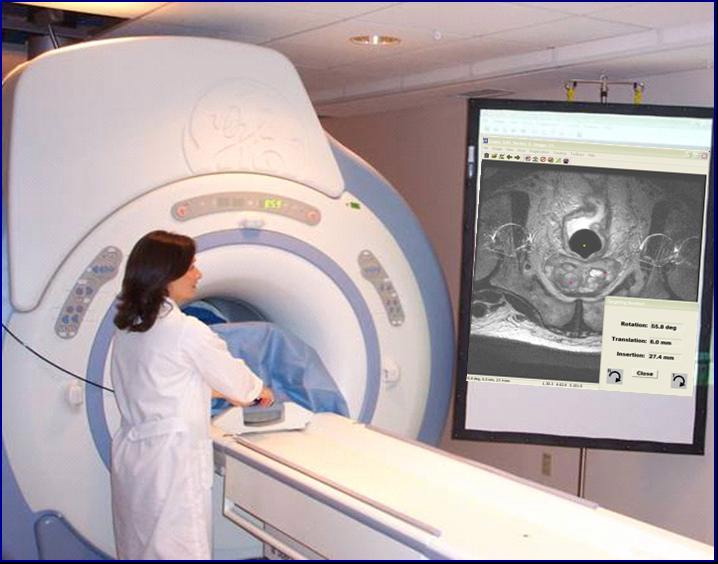
Moving for Love and MedicineA want ad, a girlfriend, and a little luck put Gabor Fichtinger on his path to medical engineering. Fichtinger, currently Associate Research Professor at the Johns Hopkins University, holds B.S. and M.S. degrees in electrical engineering and a Ph.D. in computer science from the Technical University of Budapest but he was never happy in those disciplines. In 1993, to be closer to his girlfriend – who is now his wife – Fichtinger moved to Washington, D.C., from Austin, Texas, where he'd been a postdoc at the University of Texas' Center for High Performance Computing. He found an ad in The Washington Post for a computer graphics expert for a surgical navigation team working on stereotactic radiosurgery at George Washington University. At GWU Fichtinger worked in the Division of Radiation Oncology and the Department of Radiology on computer-assisted interventional radiology and radiotherapy systems. The GWU opportunity enabled Fichtinger to find his "way home, to medical engineering." Before joining the Johns Hopkins University in 1999, Fichtinger also worked in the medical device industry, developing computer-assisted radiation therapy planning systems. "Most people get stuck in a single discipline for life, I guess. I was lucky to be able to change course several times, always adding on something new to what I had been," he says. The 45-year-old researcher's current focus is on computer-integrated surgery. An R01 grant has enabled Fichtinger's engineering team to collaborate with clinicians at the National Cancer Institute to develop a robotic system for prostate biopsy in magnetic resonance imaging (MRI) scanners. By 2025 the number of prostate cancer cases is expected to reach 500,000 a year when the bulk of baby boomers hit age 75. Fichtinger chose this area because he thought he could make a real difference in people's lives. "In prostate cancer biopsy, a modest 5% improvement may save tens of thousands of people a year," he says. Competing for and receiving the R01 grant from NIBIB in 2003 has made all the difference in Fichtinger's life as a researcher. "The R01 grant is the ultimate ticket to the grownup's table. Your name circulates. You get invited to conferences, and are asked to be on editorial boards. Suddenly you have a card in your hand," he says. "It's the ultimate cutline between anonymity and some stature in your field." One of the most important aspects of Fichtinger's academic activities is watching graduate students conceive, develop, and communicate ideas. "A good team is everything. It's priceless for the students to talk to different experts. My students are the ones who will own new methods and techniques of the future." Gabor Fichtinger is living out his dreams, but he adds "to realize your dreams you have to have the means. There is no substitute for an R01 grant."
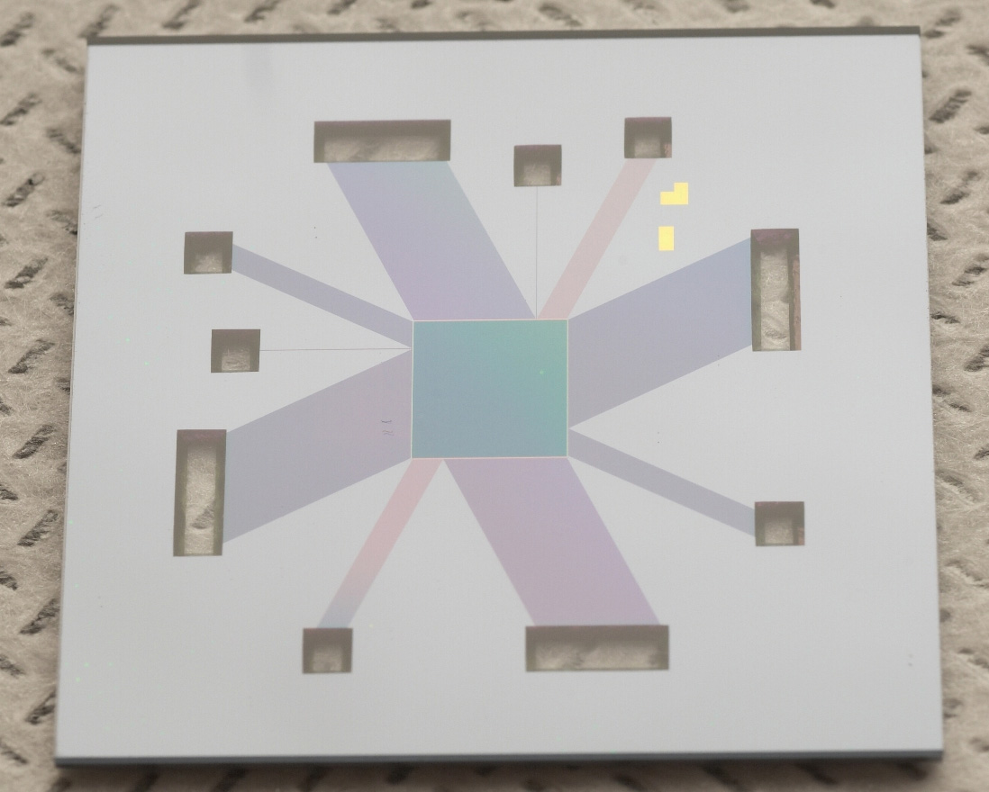
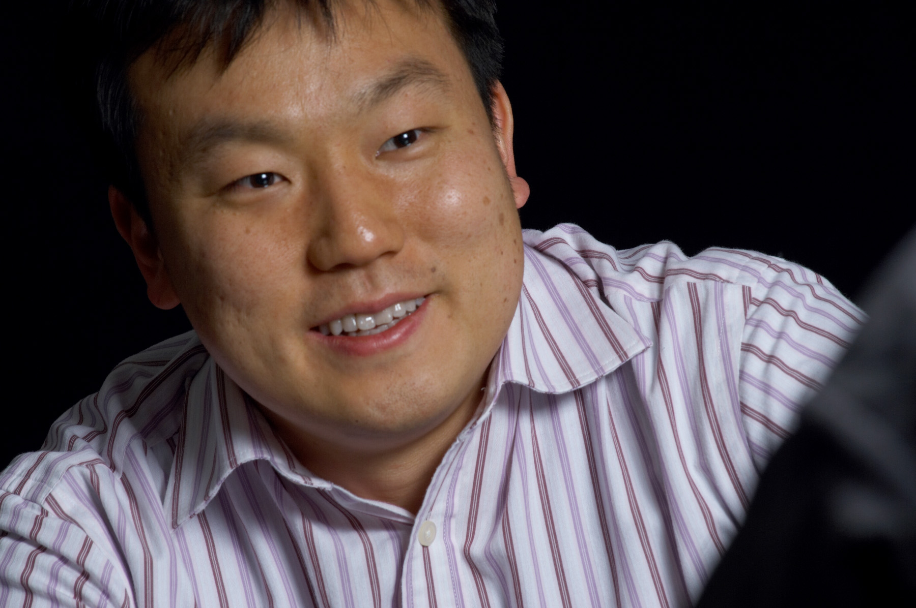
Coaxing Bright Ideas from Bright StudentsWhen Jongyoon Han arrived at Cornell University as an applied physics graduate student, he wasn't interested in biophysics and bioengineering. His Ph.D. thesis advisor, Harold Craighead, encouraged Han to try this new, emerging field in spite of Han's initial hesitation. The gentle nudging paid off because Han broke new ground with his innovative "laboratory on a chip," which can separate large DNA in minutes rather than requiring the hours typical of conventional technology. "In the end, everything worked out nicely, and I was able to make an important contribution both in physics and bioengineering because of [Craighead's] guidance and help," says Han, who earned his B.S. and M.S. degrees in the department of physics of Seoul National University, Seoul, Korea. Craighead's mentoring example made an impression on Han, who is now Karl Van Tassel Associate Professor of Electrical Engineering and an associate professor of biological engineering at the Massachusetts Institute of Technology. "I really like interacting with my students and postdocs regularly, in one-on-one meetings where we update each other on our successes and failures, as well as new ideas and directions to go," explains Han. "Many good ideas came from the discussion I had with people in my group, and I enjoy that process." Han's R01 grant to develop nanofluidic tools for proteomic sample separation has not only enabled him to develop another pioneering tool for sorting and separating biosamples, but has given him the time needed to develop his students. "The grant's 4- to 5-year timeframe enables me to bring on many very bright students and help them mature so that they can make a great impact," he says. "It's more fun to teach students because you can see them growing."Han's nanofluidics research is a long way from his master's thesis in Seoul National University – an experimental study on surface science of the Si/Au system, using a Scanning Tunneling Microscope. Says Han: "Sometimes it takes some encouragement and a bit of courage to jump into a new field. In the end, I think I was able to make the switch because I have been always interested in nanoscience and small-scale phenomena, and biology is actually a perfect example of 'working' nanotechnology."
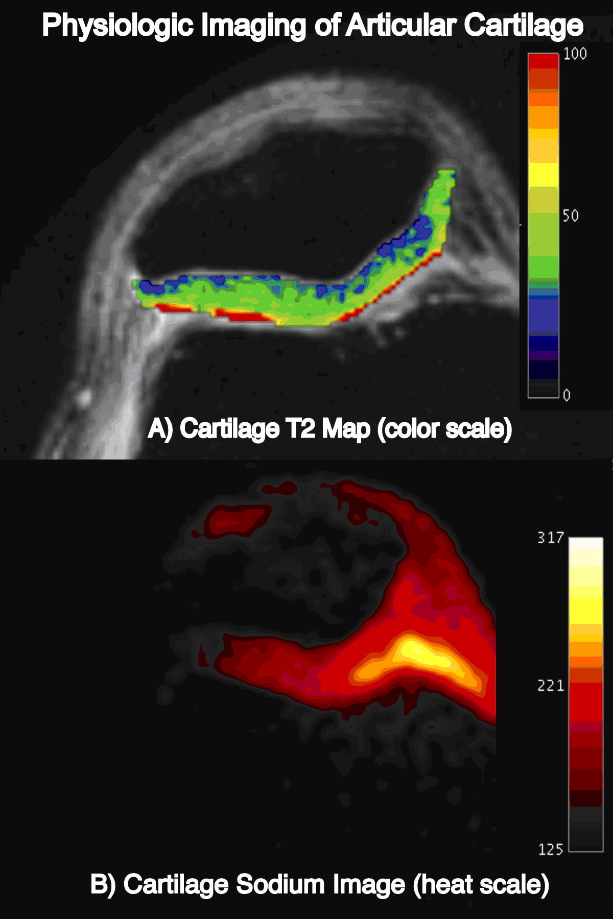
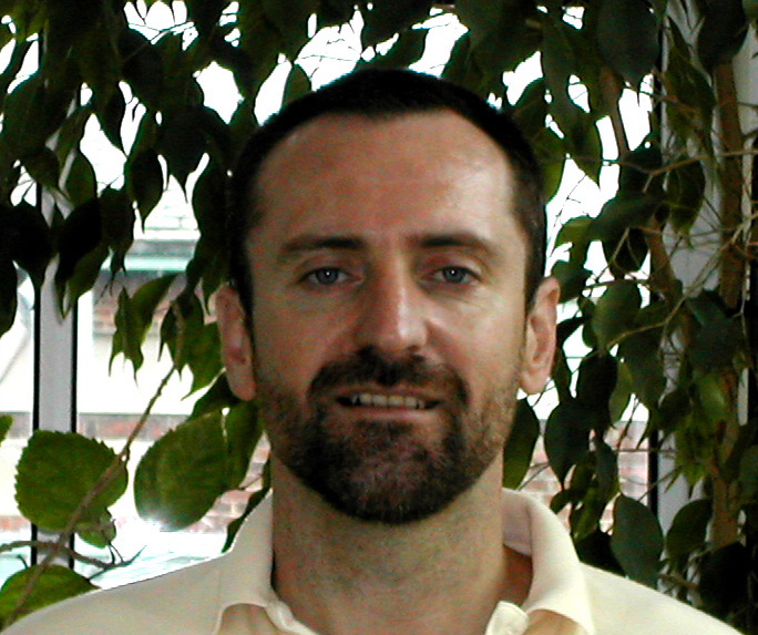
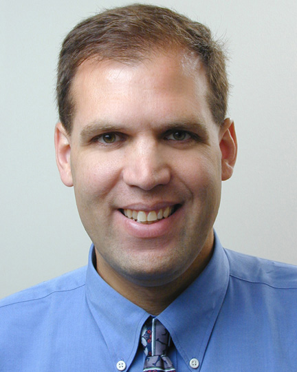
Better Clinical Medicine Builds on Strong ScienceStanford University-trained radiologist and former marathon runner Garry Gold likes to test research first hand. A few years ago, pain and swelling in a previously injured knee forced Gold to give up running. Magnetic resonance imaging (MRI) revealed a large cartilage defect. Repair involved removing a small piece of cartilage, growing it on a collagen scaffold, and reinserting the implant into the defect. While not pain free, Gold can bike, swim, and hike. To follow the implant’s progress Gold occasionally images his own knee with MRI. Definition of New Investigator - For the purpose of review and funding, applicants are considered new investigators if they have not previously served as the principal investigator (PI) on a Public Health Service supported research project other than a small grant (R03), an Academic Research Enhancement Award (R15), an exploratory/developmental grant (R21), or certain mentored research career development awards for persons at the beginning of their research career (K01, K08, K22, K23, K25, and K99/R00). Current or past recipients of Independent Scientist and other non-mentored career awards (K02, K04, K05, K24, and K26) are not considered new investigators.
|


 |
 |
Department of Health and Human Services |
 |
National Institutes of Health |
 |
