


Home > Health & Education > eAdvances
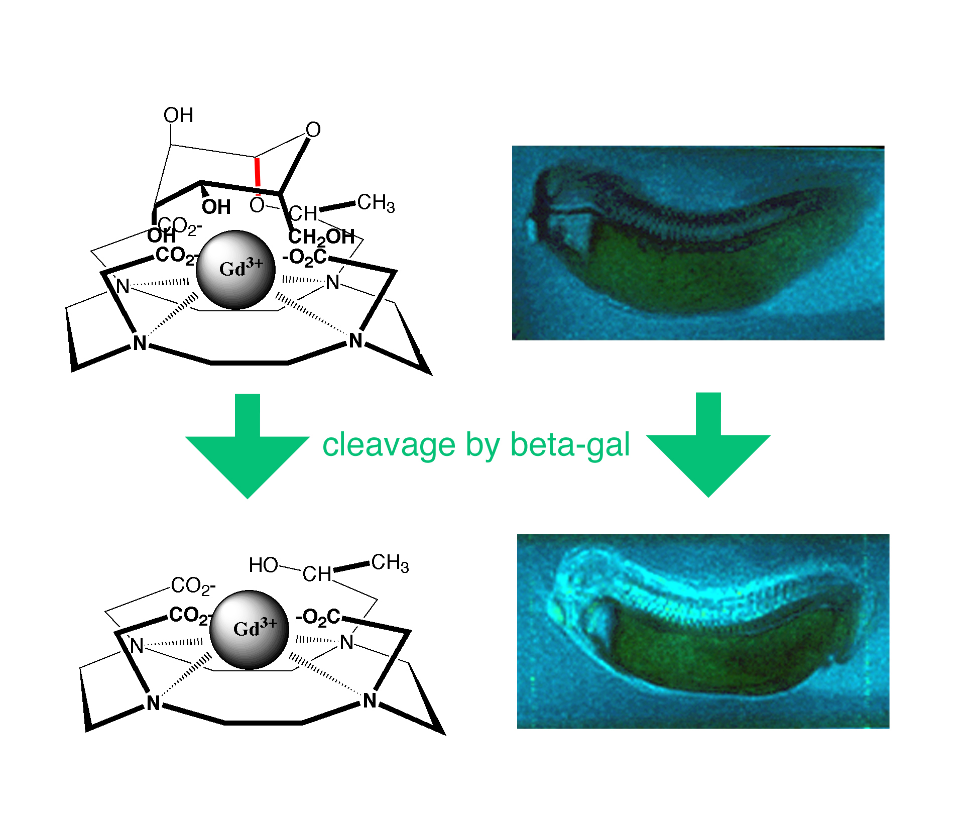
Imaging Gene Expression with Magnetic Resonance: October 23, 2006When a patient comes into the emergency room after experiencing blunt trauma, physicians need to know if any internal organs suffered damage. Magnetic resonance imaging (MRI) is widely used for this purpose. Doctors can inject an imaging dye into the patient, and then use MRI to watch the dye travel through the body. If it appears in parts of the body where it should not be, the doctor knows where the trauma is. MRI is an ideal technique for this type of imaging because it delivers three-dimensional images of the whole body, it's non-invasive and it involves no damaging radiation. But MRI only reveals anatomy, so no information is given about the metabolic processes going on inside organs or cells. What if physicians want to know if the cells are expressing genes related to damage or onset of disease? Currently, MRI can't help with this kind of question in humans, but studies by Thomas Meade, Eileen M. Foell Professor of Chemistry, at Northwestern University and his colleagues show that someday it might. Combining MR with Optical ImagingTo create a more powerful imaging modality, Meade and his colleagues are pairing MRI with optical imaging. Compared with MRI, optical imaging achieves very high resolution at the subcellular level. Scientists can image the expression of individual genes by attaching a marker gene that is activated and detected by fluorescence. When the target gene becomes active, the sample glows. If the fluorescent gene is attached to genes involved in certain types of signaling, it can alert researchers to cells that are dying or becoming cancerous. But optical imaging can only be used with individual cells or very thin slices of tissue – the signal gets hidden within anything thicker, like an organ or a whole body. By combining the strengths of MRI and optical imaging, Meade anticipates that MRI can become an important tool to study the development and molecular biology of living animals and to diagnose human diseases such as cancer or neurological disorders. Bioactivated AgentsTo enhance MRI’s ability to image cellular processes, the probes developed by Meade and his team manipulate the interaction between water molecules and the imaging dye or contrast agent. Since water molecules are present everywhere in the body, traditional MR images provide contrast that can be seen in the form of grayish hues. Meade and his co-workers have designed "smart" or bioactivated agents. They encase a contrast agent, typically a gadolinium ion, in a waterproof molecular shell. Specific enzymes can cut away the shell, allowing the water molecules to interact with the gadolinium ion and produce bright signals in a sea of grayish hues. Using this technique, Meade’s team can tell the location within the body of an enzyme as well as which gene produced it. Wherever the enzyme is, it cleaves the molecular shell and is identified in an MR image. Studying DevelopmentThe first bioactivated contrast agent that Meade and his colleagues created was a magnetic ion wrapped in a chelate capped with a sugar molecule. The target enzyme was ß-galactosidase which could cleave the sugar molecule. The researchers stitched the ß-galactosidase gene to genes of a frog and then followed the gene expression with MRI. Meade says that while ß-galactosidase would never be used in humans, it could easily be used for developmental studies in laboratory animal models. Rather than euthanizing an animal and slicing its tissues for analysis, MRI would allow researchers to see gene expression in three dimensions in a live animal. Clinical PossibilitiesSince the success of the ß-galactosidase technique, Meade and other scientists have expanded the libraries of biologically activated MR contrast agents, and they've developed several that may someday be useful in diagnosing human disease. For example, they have created MR agents that could be used to detect the expression of enzymes that become active in cancer cells, cell death, and inflammation. By detecting specific cells that are producing enzymes associated with cancer, researchers and clinicians could devise individualized treatment plans. Meade and his team have also developed contrast agents that are activated by calcium and zinc instead of by an enzyme. Calcium is critical in neural signaling, and the researchers hope that imaging calcium signaling in the brain could help detect neurological abnormalities in both adults and children. Currently, Meade and his colleagues are optimizing many of the agents they've developed. They’re examining how to make the compounds more sensitive by amplifying the number of agents on biocompatible polymers. They are also studying how to improve delivery because some of the agents do not pass easily across cell membranes or the blood-brain barrier. “We have a ways to go,” Meade says, “but there is definitely a clinical endpoint in our future." This work is funded in part by the National Institute of Biomedical Imaging and Bioengineering. References: Bull SR, Guler MO, Bras RE, Venkatasubramanian PN, Stupp SI, Meade TJ, Magnetic resonance imaging of self-assembled biomaterial scaffolds, Bioconjugate Chemistry, 16, 1323-1328, 2005.
|


 |
 |
Department of Health and Human Services |
 |
National Institutes of Health |
 |
