Home > Health & Education > eAdvances
Soft Nanotechnology Images Tumors: December 22, 2005Detection of dormant metastatic tumor cells is a critical but elusive goal in cancer treatment. To find these cells researchers are developing noninvasive optical imaging techniques that are less costly and more accessible than magnetic resonance imaging-based techniques and are free of the side effects associated with radioactive imaging agents.
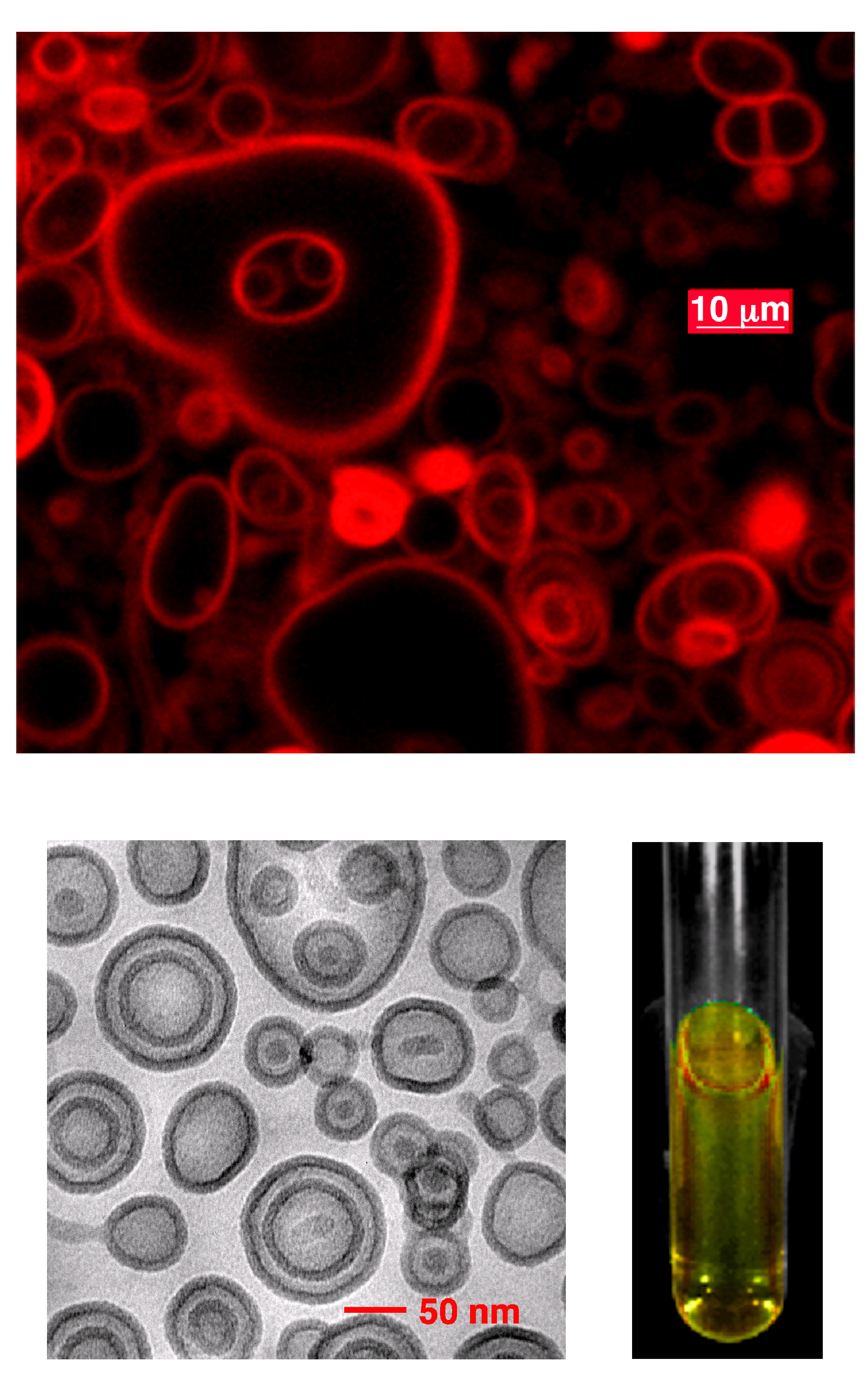
The key to the new approach is a soft form of nanotechnology called polymersomes--microscopic, flexible "bubbles" made from polymers (long-chained molecules made from repeated smaller units). A polymersome’s unique structure and material properties allow it to carry a wide range of compounds in its water resistant membrane. In a recent experiment, Peter Ghoroghchian, a graduate student in the Department of Bioengineering at the University of Pennsylvania, embedded fluorescent materials called porphyrins into the membrane of a polymersome. After injecting these emissive polymersomes directly into a tumor one centimeter beneath the surface of the skin of a mouse, he exposed the mouse to near-infrared light. The porphyrins emitted near-infrared signals detectable at the skin surface. The bright light-emitting molecules enabled a team of researchers from the University of Pennsylvania and the University of Minnesota to image the tumor. Deep Penetration with Near-Infrared LightCompounds activated by near-infrared wavelengths were chosen because near-infrared light beams are not absorbed by water, hemoglobin and other compounds that make up living tissue. Consequently, they can penetrate deeply into tissue. Other dyes used for optical imaging emit in the ultraviolet or visible portion of the optical spectrum and penetrate only about a millimeter. The large signal output of nanoscale near-infrared-emissive polymersomes will enable imaging of tissue as deep as 10 to 12 cm through breast tissue and 5 to 6 cm penetration through muscle according to Therien. "This opens up a whole array of tumors with surface activity that we could detect using this optical method--skin cancers, breast tumors, cervical cancer, and gastrointestinal cancers," says Dr. Daniel Hammer, chair of the department of Bioengineering at the University of Pennsylvania, and co-principal investigator on the project. Advantages over Other MicrobubblesPolymersomes have several potential advantages over liposomes, microbubbles which have been used for more than a decade to deliver therapeutic drugs. "Polymersomes are 20-50 times tougher. That means they can squeeze through the microcirculation and not be disrupted," Hammer notes. The mechanical strength polymersomes exhibit offers an advantage over liposomes for therapeutic drug delivery because "the contents inside the polymersome will circulate longer," he says. In addition, liposome membranes are too thin to accommodate large molecules such as optical imaging dyes called fluorophores. By choosing the length of the polymer, researchers can adjust the thickness of a polymersome’s membrane to accommodate a wide range of compounds, which will allow them to tailor the function of a polymersome to a wide range of medical applications. Polymersomes also overcome toxicity and structural issues related to quantum dots, a nonflexible infrared imaging nanotechnology made from semiconducting metals. "Near-infrared emissive polymersomes are based on materials that are nontoxic and are deformable," says Therien. Hard matter imaging agents based on quantum dots "cannot be bioresorbed, and are based on components that are known to be toxic in their elemental form. In addition, aggregation of hard matter within the circulatory system could block a capillary and lead to a stroke," he says. Targeting Cancer Cells
Clinical trials of polymersomes are still several years away, but the researchers are now focusing on targeting and imaging specific cell types with polymersomes using animal models. Ghoroghchian is currently leading an effort to make targeted versions of near-infrared-emissive polymersomes. "Cancer cells possess unique chemical markers on their surfaces," says Therien. "Cancer or inflammatory cells can be selectively imaged by attaching chemical compounds to emissive polymersomes that bind to chemical markers on surfaces of these cells. Because the amplitude of the emissive signal emanating from targeted near-infrared-emissive polymersomes is directly related to the number of cancer cells present, this optically-based imaging approach provides the physician with quantitative information impossible to obtain with techniques such as magnetic resonance imaging." This project would aid in detecting small tumors or dormant or latent metastases that involve only a small number of cells. To improve tumor staging and classification, the researchers are developing polymersomes that characterize how a tumor changes over time. By formulating fluorophores to emit at different wavelengths, the researchers can create a family of polymersomes that target various receptors on a tumor’s surface. "These receptors provide an index of how the tumor has mutated or evolved," says Hammer. A clearer picture of the developing tumor "would allow a clinician to determine the best line of treatment for that particular strain of tumor," he adds. Also in development are polymersomes that would deliver chemotherapy agents directly to a tumor. The surface of the polymersomes would carry a molecule that would bind to tumor cells, its membrane would disperse fluorophores for optical imaging, with the chemotherapy "payload" carried in the interior. Currently, researchers are designing a mechanism that would allow an external stimulus, such as exposure to light or a sudden change in temperature, to destroy the polymersome membrane, releasing the therapeutic drug. If successful, this approach would allow clinicians to deliver chemotherapy in a much more targeted manner: Optical imaging would allow them to confirm that polymersomes have reached and bound to the target tumor, while the external stimulus would trigger release of the chemotherapy agent directly to the tumor. These synthetic but biocompatible nanoparticles offer a new mode of transport for optical imaging agents as well as an improved method of disease surveillance. "We’re excited about near-infrared-emissive polymersomes because they are so incredibly versatile," says Hammer. "Their material properties can be designed for the specific task for which they’re going to be used. We think this represents a new paradigm that will be applicable to many different medical applications." Funding for the research was provided by the National Institute of Biomedical Imaging and Bioengineering. Reference: Ghoroghchian P et al., Near-infrared-emissive polymersomes: Self-assembled soft matter for in vivo optical imaging, Proceedings of the National Academy of Sciences, 102, 2922-2927, 2005.
|
 |
 |
Department of Health and Human Services |
 |
National Institutes of Health |
 |


.jpg)