


Home > Health & Education > eAdvances
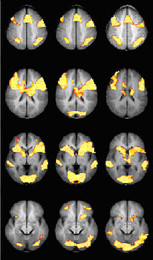
Medication Boosts Brain Activity in Kids With Attention, Reading Disorders: August 11, 2005Using a sensitive new functional magnetic resonance imaging (fMRI) technique, scientists have identified a brain region that underfunctions in adolescents with attention deficit hyperactivity disorder (ADHD) or a reading disorder. In addition, the researchers found, a standard treatment using the drug methylphenidate causes that brain region to become more strongly activated.ADHD affects three to five percent of American school-aged children, with symptoms that include an inability to restrain impulsive behavior or to pay attention for long periods of time. These symptoms, often lasting into adulthood, are associated with lower achievement levels in school and at work. The signaling pathways in the brain that are involved in ADHD have come under intensive study in recent years. But most of this research has focused on impulsivity. Inattention has been more difficult to study, partly because the neural circuitry of attention extends throughout different brain regions. “A number of distributed systems are responsible for attention,” says Dr. John Gore, director of the Institute for Imaging Science at Vanderbilt University. Collaborator Dr. Keith Shafritz, of the Center for Brain Imaging and Analysis at Duke University, adds, “We wanted to find out what role each particular brain region plays in attentional circuitry.” ADHD offers a chance to look closely at the attention-related regions one by one. Tracing a picture of how the brain functions when people focus or shift their attention requires a technique for detecting subtle shifts in activity patterns. With the help of a grant from the National Institute of Biomedical Imaging and Bioengineering, Drs. Gore and Shafritz developed a specialized and sensitive method of analyzing fMRI scans of the brain. Although the use of fMRI to examine ADHD is not new, this recent research is innovative in several respects. It is the first double-blind, placebo-controlled study to look at the effects of methylphenidate on brain activity. “This is a more stringent type of study,” says Dr. Shafritz. “We didn’t know and the children didn’t know whether they were taking the real medication or a placebo, so their subjective opinions could not influence the results.” This was also the first neuroimaging study to compare the brain activity of ADHD subjects to that of people with another, similar disorder—in this case a reading disorder. Also, says Dr. Gore, “This is one of very few fMRI studies to look at a psychoactive drug to see what areas it affects during a particular mental task.” More commonly, studies look at how a drug affects the outcome of a particular task, rather than how the results are produced. The main goal of the study was to discover which brain regions function abnormally both in individuals with ADHD and in individuals with a reading disorder. The two disorders often occur together. The study population comprised four groups: 15 adolescents with ADHD alone, 8 with reading disorder alone, 4 with both reading disorder and ADHD, and a control group of 14 who had neither disorder. Except for those in the control group, each person underwent two brain scans while performing two attention-related tasks—one after receiving a dose of methylphenidate and one after receiving a placebo. In the unmedicated portion of the experiment, the subjects with ADHD or reading disorder had significantly less activity than the control subjects in a part of the brain known as the striatum. After they received methylphenidate, however, their activity in this brain area rose to match that of the control group, but their performance on the attention tasks did not improve. “This is a surprising finding,” says Dr. Shafritz. “We had expected that an increase in activation would equal an increase in performance, but we found no correlation.” These results leave the scientists with scant answers but plenty of room for more investigation. One new question is why ADHD and reading disorder, which affect children in two different ways, yield fMRI scans that show similar brain dysfunctions and a similar response to medication. Another topic to explore is the primary effect of methylphenidate. Does it moderate impulsivity and hyperactivity more than it bolsters attention? With Duke psychologist Dr. Jeff Epstein, Dr. Shafritz is now developing a study to look at potential differences in brain response to different drug treatments for ADHD. He expects that study to open yet more questions. Besides support from NIBIB, the scientists received funding from the National Institute of Mental Health and the National Institute of Child Health and Human Development. Reference: Shafritz KM, et al., The effects of methylphenidate on neural systems of attention in attention deficit hyperactivity disorder. American Journal of Psychiatry 161:1990-1997, 2004. |


 |
 |
Department of Health and Human Services |
 |
National Institutes of Health |
 |
