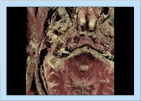
High Resolution MRI of Auditory Canal [23 seconds]
This movie shows the raw image data collected from a high resolution 3T MRI scan of the head with zoom-in viewing of the middle ear, showing the semicircular canals and cochlea and other internal head structures. Richard A. Robb, Ph.D.
c. 2006 Mayo Clinic College of Medicine
Localized Clot Formation [1 minute 4 seconds]
This two-photon fluorescence image sequence shows the formation of a localized photothrombotic clot in a surface arteriole. The fluctuating, streaked appearance of the vessels is due to the motion of red blood cells and indicates flow. A saturated white strip at the top of the frame indicates irradiation with 523-nm light. Clot material is formed just downstream from the irradiated region. David Kleinfeld
University of California, San Diego
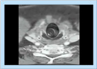
Quantitative Virtual Colonoscopy [33 seconds]
This movie shows a 3-D CT volume scan of the colon rendered into a virtual colonoscopy fly-through to visualize internal colonic structures, such as polyps. Further processing of this data reveals the microvasculature within the colon lumen and any polyps present during the virtual endoscopic fly-through. Microvessel density is a prediction of metastatic potential. Richard A. Robb, Ph.D.
c. 2006 Mayo Clinic College of Medicine
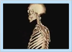
Skeleton from Helical CT Scan [29 seconds]
Body volume CT scans can be processed to reveal the internal bone structures from head to foot. This movie shows the skeleton of a subject scanned with such a procedure. Richard A. Robb, Ph.D.
c. 2006 Mayo Clinic College of Medicine
Virtual Bronchoscopy [19 seconds]
This movie shows the luminal surface of the branching bronchi, which can be visualized down to several orders of branching. The surfaces of the airways can be rendered translucent to see through them for examination of adjacent tissues external to the pulmonary airways. Richard A. Robb, Ph.D.
c. 2006 Mayo Clinic College of Medicine
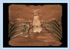
Virtual Cardiac Endoscopy [29 seconds]
Rapid volume CT scans of the chest can be processed to produce endoscopic fly-throughs through the chest into the heart cavities. The viewpoint moves from outside of the chest moving into a pulmonary vessel and proceeding into the left atrial chamber and back out again. Richard A. Robb, Ph.D.
c. 2006 Mayo Clinic College of Medicine