


|
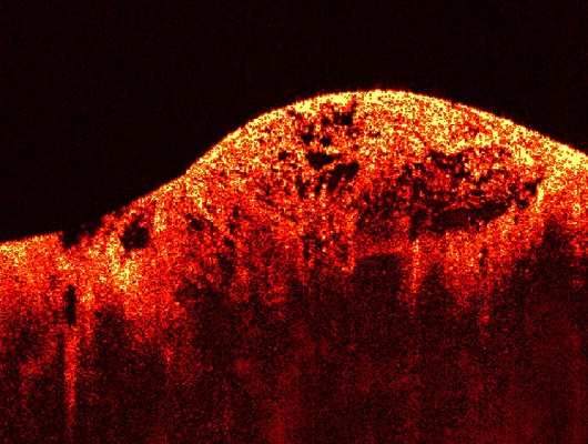
Optical Coherence Tomography (OCT) image of a sarcoma This image of a sarcoma, or muscle tumor, was obtained using Optical Coherence Tomography (OCT). This technique is similar to ultrasound, and yields images of tissues below the surface. The images are on a micron scale, and they are gathered by measuring scattered light waves that bounce off of tissue. In this picture, the tissue looks healthy and normal on the left. To the right, the structure appears cancerous and irregular. Images like this one, obtained by using OCT in real-time, can help to promote human health by detecting tumors early during image-guided procedures, and allowing time for treatment. Image courtesy of Dr. Stephen Boppart, Biophotonics Imaging Laboratory, University of Illinois at Urbana-Champaign. EB 005221
|

 |
 |
Department of Health and Human Services |
 |
National Institutes of Health |
 |
