The Whole Ball of Wax: TB's Distinctive Cell Wall
 |
| Patrick Brennan, Ph.D. |
In 1882, Robert Koch had to invent a two-step staining procedure to get Mycobacterium tuberculosis (M. tb) to show up under his microscope lens. He speculated on why M. tb was so difficult to stain: "It seems likely that the tubercle bacillus is surrounded with a special wall of unusual properties." Unusual is an understatement: The waxy coats of mycobacteria (including the microbes responsible for TB and leprosy) are unique among living things.
Patrick Brennan, Ph.D., of Colorado State University, uses words like "amazing" and "chemically extraordinary" to describe M. tb's cell wall. Combining chemical and genetic techniques, he discovered some of the methods M. tb uses to make its wall. Continued research may lead to drugs that can squelch the microbe's ability to build up its wall and thus make it more vulnerable to destruction.
Chicken Wire and a Sugar Coat
 |
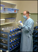
TB bacteria grown in large quantities
Credit: Cell culture laboratory—John Belisle, Colorado State University |
Bacterial cell walls, explains Dr. Brennan, have a layer of chicken wire-shaped molecules (peptidoglycan) that give the wall rigidity and enclose the microbe's inner workings. Penicillin can kill certain bacteria by snipping apart the chicken wire. M. tb, too, has a layer of these molecules overlaying its innermost cell membrane. However, the TB microbe has three more layers that further insulate it from attack. Atop the chicken wire is a sugary coating (arabino-galactan) that forms a bridge to the third layer, which is packed with stringy molecules called fatty acids. Many kinds of cells contain fatty acids, but M. tb's (called mycolic acids) are exceptionally long. The tangle of mycolic acids is wrapped in a final layer of tightly packed waxy molecules that make the cell nearly waterproof.
Beginning in the 1940s, chemists studied M. tb's cell wall by grinding up cells and extracting the assorted components for further analysis. It was not until the 1980s and the development of tools such as nuclear magnetic resonance and mass spectrometry, however, that chemists could determine the molecules' shapes, the chemical steps in their manufacture, and how they are arranged in an intact cell. In 1998, came a critical breakthrough—determination of M. tb's genetic sequence. Researchers gained clues into the genetic reasons behind M. tb's structural anomalies and its uncanny ability to survive in the human lung for long periods.
With the entire gene sequence in hand, Dr. Brennan and his colleagues started to tease out which genes M. tb must have to build its rugged cell wall. Those essential genes encode enzymes and they, in turn, might fall prey to specifically designed drugs, explains Dr. Brennan. In October 1990, the team at Colorado State University developed a more detailed picture of the multilayered cell wall. The core cell wall, they learned, is enormous—the biggest bacterial macromolecule ever discovered. This immense molecule has an equally long name that links together all of its components: mycolyl-arabino-galactan-peptidoglycan, or mAGP.
Breaking Down the Wall
Aided by a better understanding of the enzymes M. tb needs to make its wall, Dr. Brennan and Gurdyal Besra, Ph.D., (then a postdoctoral student in Dr. Brennan's lab), took a closer look at a drug called TLM. Through genetic engineering, the researchers created strains of mycobacteria that produced an overabundance of two key enzymes needed for the first steps in mycolic acid manufacture. Although TLM readily kills normal M. tb, it had little effect on the mutants. The conclusion: TLM targets and disrupts one or both of these required enzymes, thus inhibiting mycolic acid formation.
Refinements to the picture of M. tb's cell wall construction continue to be made with an eye towards finding drugs capable of destroying the microbe's wall. Among the efforts is an NIAID-funded consortium joining researchers from NIAID's intramural program, Colorado State University, St. Jude Children's Research Hospital in Tennessee, GlaxoSmithKline in Pennsylvania, and the University of Newcastle Upon Tyne in England.
 |
| Schematic of the bacterial cell wall. Credit: Michael Niederweis, Universität Erlangen, Germany |
back to top