Our Science – Emmert-Buck Website
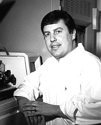
Michael R. Emmert-Buck, M.D., Ph.D.
 |
|
|||||||||||||||||||||
Biography
Dr. Emmert-Buck received his M.D. and Ph.D. degrees from Wayne State University School of Medicine. His clinical training in anatomic pathology was conducted in the Laboratory of Pathology, NCI, and completed in 1995. In 1996, he was promoted to unit chief to head the Pathogenetics initiative.
Research
The Pathogenetics Unit develops new approaches to investigate the molecular pathology of cancer. These methods are subsequently applied to gene discovery projects, often as collaborative, multidisciplinary efforts with investigators in the NIH intramural program.
We are currently working on developing two new technologies; expression microdissection (xMD) and layered expression scanning (LES). Based on immuno-targeting, xMD converts microdissection from an operator-dependent to an operator-independent mode, thus eliminating the need to laboriously procure cells. When fully developed xMD likely will offer several advantages over current methods, including; a) increased dissection rate (several orders of magnitude), b) increased dissection precision (subcellular), c) removal of variance among individual operators, permitting standardization of the dissection process, and d) elimination of targeting difficulties due to poor image quality of non-coverslipped (non-index-matched) histology sections used in current dissection systems.
Layered expression scanning (LES) is a new technology co-developed by the Pathogenetics Unit and 20/20 GeneSystems, Inc. through a Cooperative Research and Development Agreement (CRADA). The method utilizes a layered array of membranes for high-throughput molecular analysis and can be applied to a variety of life science platforms, including tissue sections, cells in culture, electrophoresis gels, multi-well plates, and tissue arrays. The technique is performed by passing the sample through a series of membrane layers while maintaining the original two-dimensional architecture. Each membrane measures a different molecular species, thus multiple RNA transcripts or proteins in each sample element (e.g., various cellular phenotypes in a tissue section, bands on a gel, individual wells of a microtiter plate) can be simultaneously analyzed. The method is conceptually simple, requires no moving parts, and can be used as an open or closed format.
Laser Capture Microdissection (LCM) was co-developed by the Pathogenetics Unit along with Drs. Lance Liotta and Robert Bonner. Commercialization of a laser-based microdissection instrument was successfully completed in 2001 via a CRADA with Arcturus Engineering. LCM enables investigators to specifically dissect cells from a section of complex tissue under direct microscopic visualization. The method entails placing a thin transparent film over a section and selectively adhering targeted cells of interest to the film with a fixed-position, short-duration pulse from an infrared laser. The film is then removed and the biomolecules from the procured cells are ready for molecular analysis. LCM technology also provides experiment documentation, as dissection occurs in real time and can be imaged on the PixCell and AutoPix systems.
Overall, we anticipate that xMD, LES, and LCM will become complementary tools that will assist the Unit and other investigators in phenotype- and expression-based profiling studies of cell populations in tissue sections.
As part of an integrated effort between the Pathogenetics Unit and the Urologic Oncology Branch, CCR, we are applying both standard and new methodologies to determine the molecular profiles associated with progression of prostate cancer. For example, using LCM and an expression array approach, a set of 21 genes were identified that segregate high- and moderate-grade prostate cancer. The biological significance and potential diagnostic utility of this gene set are being examined in the laboratory.
In a search for genes involved in the development of esophageal tumors, we are collaborating with the Cancer Prevention Studies Branch, CCR, to examine the molecular basis of esophageal squamous cell carcinoma (ESCC) in a high-risk population in Shanxi province in China. Allelotype comparison of patients with and without a family history of ESCC identified a region on chromosome band 13q12 that may harbor a familial tumor suppressor gene.
Multiple Endocrine Neoplasia Type I (MEN1) is an inherited syndrome characterized by development of neuroendocrine tumors in affected individuals. The responsible gene was discovered in 1997 by the NIH MEN1 working group, including the Pathogenetics Unit and groups from the National Human Genome Research Institute, the National Center for Biotechnology Information, and the National Institute of Diabetes and Digestive and Kidney Diseases. Current efforts of the Pathogenetics Unit include characterization of MEN1 gene mutations in neuroendocrine tumors, and immunohistochemical evaluation of menin in human tissues.
This page was last updated on 6/11/2008.

