Contents
- Critical Resources
The Operating Room of the Future
Critical Resources
The Operating Room of the Future
Advanced imaging technologies are transforming therapy.
By Laura Bonetta
When you visit a hospital today, the most low-tech place is the operating room," says Ferenc Jolesz, director of the National Center for Image-Guided Therapy (NCIGT) at Brigham and Women's Hospital and Harvard Medical School in Boston. "Advanced technologies are available for diagnosis but not for therapy. That is at the heart of what we are trying to do."
The NCRR-supported National Center for Image-Guided Therapy (NCIGT) at Brigham and Women's Hospital and Harvard Medical School in Boston will soon unveil the first prototype of its "operating room of the future." This will comprise several imaging systems, a sophisticated surgical table that moves patients between stations, and detailed visual displays to guide a clinician during medical procedures. By developing approaches that incorporate powerful imaging technologies in the operating room, NCIGT researchers will assist clinicians in delivering safer and more effective treatments.
The center is one of 50 NCRR-funded Biomedical Technology Research Resources nationwide, each focused on developing advanced technologies for biomedical research and clinical practice. With co-funding from the National Cancer Institute and the National Institute of Biomedical Imaging and Bioengineering, NCIGT serves as a test bed for new imaging technologies and their application in the operating room, where they can assist surgeons in delivering safer and more effective treatments.
"In traditional surgery, the surgeon's vision is limited to the surface," Jolesz says. "With imaging, you can see every layer of tissue without having to cut through the patient." New imaging technologies allow surgeons to "see" where tumors are located, for example, even before starting an operation. Imaging-based approaches also enable new treatment methods that do not require any cutting at all. And imaging methods allow medicines to be delivered to the intended target with greater precision and effectiveness.
NCIGT's accomplishments in the past decade illustrate both the promise and the challenges of bringing advanced technologies from the laboratory to the clinic. They also reveal the key ingredients for making this transition possible: a strong research tradition, teams of basic scientists and physicians focused on translating a research finding to a particular application, and collaborations among different research institutions and between academic researchers and industry.
BIOMEDICAL TECHNOLOGY RESEARCH RESOURCES FOR IMAGING TECHNOLOGIES
Twelve of the 50 NCRR-funded Biomedical Technology Research Resources (BTRRs) focus on pursuing cutting-edge development and improvement of methodologies and technologies for imaging and spectroscopy. These technologies are used to study organ structure and function, perfusion, and oxygen extraction and metabolism for the diagnosis, staging, treatment, evaluation, and investigation of diseases and abnormalities. The imaging centers are located in major research-intensive medical centers, providing a nurturing environment for interdisciplinary research and exceptional opportunities to identify proper collaborations to drive the development of technology. This proximity contributes as well to the reduction in translation barriers to clinical applications
BioCurrents Research Center — development of tools to follow the dynamic properties of living cells. Principal Investigator: Peter J.S. Smith, Ph.D.
Center for Advanced Magnetic Resonance Technology at Stanford — development of magnetic MRI techniques in humans and animals. Principal Investigator: Gary H. Glover, Ph.D.
Center for Functional Imaging Technologies — development of neuroimaging techniques in humans. Principal Investigator: Bruce R. Rosen, M.D., Ph.D.
Integrated Center for In Vivo Microscopy — development of techniques for very high-resolution imaging of small animal models. Principal Investigator: G. Allan Johnson, Ph.D.
National Center for Image-Guided Therapy — image-guided therapy. Principal Investigator: Ferenc A. Jolesz, M.D.
National Center for Microscopy and Imaging Research — development of tools and techniques for electron microscopy at the cellular level. Principal Investigator: Mark H. Ellisman, Ph.D.
National Center for X-Ray Tomography — X-ray microscopy at the cellular level. Principal Investigator: Carolyn Larabell, Ph.D.
Neuroimage Analysis Center — Understanding the human brain through imaging. Principal Investigator: Ron Kikinis, Ph.D.
NMR Imaging and Localized Spectroscopy — MRI using ultra-high magnetic fields. Principal Investigator: Kamil Ugurbil, Ph.D.
Resource for Magnetic Resonance and Optical Imaging — high-performance computing. Principal Investigator: John S. Leigh, Ph.D.
Resource for Quantitative Functional MRI — development of MRI techniques in humans. Principal Investigator: Peter C.M. van Zijl, Ph.D.
Southwestern NMR Center for In Vivo Metabolism — imaging techniques to understand metabolic changes in humans. Principal Investigator: Craig R. Malloy, M.D.
For more information about these and other technologies supported at BTRRs, visit www.ncrr.nih.gov/BTRR.
Ultrasound as a scalpel
A technology that progressed rapidly from discovery to clinical implementation is magnetic resonance–guided focused ultrasound. Physicians typically use ultrasound to image tissues in the body (see "An Imaging Primer"); when high-frequency sound waves bounce off internal tissues, the echoes they produce can be analyzed as images of tissues. However, it has been known for more than 60 years that focusing a beam of ultrasound to a particular point in the body could be used to destroy or remove a tissue — akin to performing surgery but without a scalpel.
In the early 1990s, Kullervo Hynynen and Jolesz, both at Brigham and Women's Hospital at the time, showed that this type of "surgery" could be performed inside a magnetic resonance imaging (MRI) scanner to guide and monitor the procedure. Use of the MRI allows surgeons to carefully plan where to point the ultrasound and to immediately visualize the results of the operation. In addition, because energy from the ultrasound beam produces heat, a special application of MRI allows surgeons to monitor the temperature in the target tissue and maintain it within a certain range.
Hynynen and Jolesz have taken their research to the clinic. Research by the Focused Ultrasound Laboratory core at NCIGT led to the development of an instrument, called ExAblate 2000, for transmitting focused ultrasound waves. This instrument was approved by the U.S. Food and Drug Administration (FDA) in 2004 for the removal of fibroids.
Fibroids are generally benign muscular tumors that grow in the wall of the uterus. Most women with fibroids do not have symptoms, but for those whose fibroids cause pain or severe bleeding, removal of the uterus is one of the few options available to them. But with focused ultrasound, a fibroid can be removed without surgery.
A woman lies facedown on the MRI scanner, and beneath her the ExAblate 2000 ultrasound transducer transmits a focused stream of waves for up to several hours. The heat generated by these waves gradually destroys the fibroid, while the MRI scanner continuously monitors the temperature of tissues in the uterus as well as the position of the fibroid. The recovery time for the patient is reduced, and she is able to return to normal activities more quickly than after traditional surgery.
Because of the success of focused ultrasound with uterine fibroids, NCIGT researchers are testing the same technology on different organs, such as the prostate or brain, by modifying the transducer and how the treatment is delivered. These procedures are at various stages of testing in animals or in clinical trials in patients.
Focused ultrasound is also proving useful for the delivery of medicines to the brain. Humans have a collection of tightly packed epithelial cells within the blood vessels leading to the brain — called the blood-brain barrier — which prevents most larger molecules, including drugs, from passing through the bloodstream into the brain. Researchers in the Focused Ultrasound Laboratory core have developed a technology that uses focused ultrasound to poke temporary holes in the blood-brain barrier to allow drugs through. So far, the strategy has been successful in delivering chemotherapy for brain tumors in animal models.
AN IMAGING PRIMER

Functional magnetic resonance imaging reveals areas of the brain that become active when an individual performs a particular task. This type of imaging is used to create maps of human brain function.
Magnetic resonance imaging (MRI) uses a powerful magnetic field and pulses of radio wave energy to construct images of the structures and organs inside the body. MRI can provide a greater level of detail for some areas of the body than can other imaging systems. It is especially useful in neurological, musculoskeletal, cardiovascular, and oncological imaging. The strength of the magnetic field in an MRI scanner is measured in Tesla units. The standard MRI systems used for patient care imaging are 1.5 Tesla. But more powerful and faster 3-Tesla MRI systems are becoming more common in hospitals and research institutions.
X-rays are a form of ionizing radiation that can travel through the body. When X-rays strike a film, they produce a picture. Dense tissues in the body, such as bones, absorb many of the X-rays and appear white on the film, whereas less dense structures appear as different shades of gray.
Functional MRI (fMRI) measures signal changes in the brain due to changing activity. Increased neural activity causes an increased demand for oxygen and thus an increase in the amount of oxygenated hemoglobin relative to deoxygenated hemoglobin, which can be detected as a stronger MR signal.
Computed tomography (CT) uses X-rays and computer processing to generate a three-dimensional image of the inside of the body from a series of images taken from different angles. CT scanning is a good tool for examining bone and calcifications within the body or such structures as blood vessels.
Ultrasound consists of high-frequency sound waves that can produce images when they reflect off organs and other structures in the body. The most well-known imaging application for ultrasound is producing pictures of fetuses in the womb.
Positron emission tomography (PET ) detects gamma rays emitted by a positron-emitting radionuclide (tracer) ingested by a patient. Images of tracer concentration in three-dimensional space within the body are reconstructed by computer analysis. The reconstruction is typically accomplished with the aid of a CT scan performed on the patient during the same session in the same machine.
Forging new paths
The first step in applying any technology to a clinical area, such as prostate or brain tumor treatment, is to assess whether there is a need for it. "There has to be a clinical need to do something better than the current treatment," Jolesz explains. The second step is to look for a technology that can provide the needed improvement. "Technology is never the initiating process. We don't develop a technology and then look for an application."
NCIGT researchers benefit from working at a hospital and having access to and interactions with physicians who can both communicate their needs and provide feedback on new technologies. "When you are using imaging in either research or diagnosis, it is like reading pages in a book," Jolesz says. "But when you use it in therapy, the imaging system has to change in relation to what the surgeon is doing. It becomes an interactive system, so the requirements are different."
But the application of new technologies to the clinic doesn't require just collaboration with physicians. Usually new tools and instruments are needed, and researchers must work closely with industry to develop them. "We might have an existing technology available for a diagnostic system but need new features to make it suitable for surgery," Jolesz says. "You need a company to make the devices and components. They are complex and expensive and cannot be developed in our center."
The ExAblate 2000 ultrasound device, for example, was built by InSightec, a company based in Haifa, Israel. General Electric developed the MRI device used to image the uterus while ultrasound energy is applied to fibroids. "We are constantly meeting with people from different companies," Jolesz says, adding that for some applications, as many as 100 distinct companies may become involved at different stages of development.

During a surgical procedure, a clinician not only views the organ being operated on but also has access to digital displays of a variety of images obtained before or during the procedure. For example, several imaging technologies can be combined to provide neurosurgeons with unparalleled views of physical structures and functional areas of the brain. The images, such as the one shown to the right of the photograph, serve as roadmaps to guide a surgeon throughout a procedure.
Sometimes companies do not step forward to develop the needed tools. For example, NCIGT researchers developed a technology to improve the treatment of cardiac arrhythmia, a form of heart disease caused by abnormally fast or unusually slow heart rates, but the researchers are missing a crucial piece. To treat some types of cardiac arrhythmias, a physician guides a catheter with an electrode at its tip to the area of heart muscle at which there is abnormal electrical activity. The catheter is typically guided by X-ray imaging, but NCIGT researchers have developed a technology that uses more powerful and accurate MRI. "We need new types of catheters that are MR compatible, but there are no products available," Jolesz says. "Without them, the technique is not going to work. So we have to wait."
Collaborations also are ongoing between NCIGT researchers and researchers at other institutions and organizations. One such example is a collaboration to try to make surgery guided by powerful MRI instruments more widely used.
The collaboration involves Clare Tempany, who codirects NCIGT with Jolesz and heads the Image-Guided Prostate Therapy core. Over the past decade, she has pioneered many new procedures and tools to perform prostate biopsy and therapy under the guidance of MRI. Therapy for prostate cancer is often administered using "seeds," small radioactive rods implanted directly into the tumor and prostate gland. The seeds are very powerful but only deliver radiation within a few millimeters of where they are implanted, so their placement is of critical importance. Tempany and colleagues have developed MRI-based methods of imaging the tumor, mechanically controlling the needle that carries the seeds, and recognizing the placement of the needle. Now they are trying to adapt these same procedures to much more powerful, high-field MRI scanners, such as those that are 3 Tesla — the unit used to measure magnetic field — in field strength. During a surgical procedure, a clinician not only views the organ being operated on but also has access to digital displays of a variety of images obtained before or during the procedure. For example, several imaging technologies can be combined to provide neurosurgeons with unparalleled views of physical structures and functional areas of the brain. The images, such as the one shown to the right of the photograph, serve as roadmaps to guide a surgeon throughout a procedure.
IT ALL STARTED WITH THE BRAIN
The development of many imaging approaches now applied to different diseases and conditions was initially driven by a need in neurosurgery. The brain is enclosed in a bony box, making it difficult for the surgeon to visualize various structures, and yet more than in any other organ of the body, surgeons need to know precisely where and what they are cutting. "It is critical to preserve any healthy tissue," says Alexandra Golby, head of NCIGT's Image-Guided Neurosurgery core. "You cannot take a little bit extra because that could lead to lifelong deficits."
Thanks to advances in several types of imaging technologies over the past 100 years, neurosurgeons today have access to images of the main structures, or landmarks, in a patient's brain prior to an operation — like having a roadmap before going on a trip. Golby has been working to add "function" to the map.
“Not all areas of the brain are equally important,” she says. “For example, some areas are critical for motor and visual skills. But no labels in the brain read ‘Don’t cut here.’” To determine where these critical areas are, Golby uses functional MRI, a technique that detects blood flow changes in the brain, indicating brain activity. “Functional MRI is becoming slowly accepted as a clinical tool for presurgical mapping,” Golby says. “It is a technically demanding procedure. What we are trying to do now is to make it more turnkey.”
Another imaging technique that adds new landmarks to the preoperative map is diffusion tensor imaging, a variation of MRI that detects connections between different brain areas. "We are trying to combine as much information as possible," Golby says. "The goal is to get as complete a picture as possible." In addition to taking images before an operation, NCIGT researchers have pioneered the use of MRI to obtain pictures of the brain during an operation, or intraoperatively. "During surgery, the brain shifts due to many factors, including resection of tissue, making preoperatively acquired information progressively less useful," Golby explains. "So intraoperative imaging helps the surgeon know where the limits of resection should be."
But how can all these pre- and intraoperative imaging data fit onto the surgeon’s roadmap? This is where bioinformatics and computer programming play a part. In collaboration with researchers at the Center for Integration of Medicine and Innovative Technology, another NCRR-funded Biomedical Technology Research Resource, and the NCRR-funded Biomedical Informatics Research Network (BIRN), NCIGT scientists have developed computer programs and algorithms to analyze and integrate different imaging data. More information about BIRN
The software package 3D-Slicer (freely available to all researchers at www.slicer.org) is widely used to construct and visualize collections of MRI data. With Slicer, the surgeon can manipulate the data to obtain images of the brain from different angles and zoom in at different sites. In addition to producing 3-D models from conventional MRI images, Slicer presents information derived from functional MRI, diffusion tensor imaging, and electrocardiography. Researchers are now adapting Slicer to other applications. "Neuroimaging is very sophisticated, but many of the same techniques are now being adopted in other areas, such as prostate surgery," Tempany says.
LOOKING FORWARD
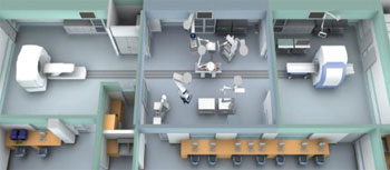
Many of the technologies used to image the body before and during surgery are becoming components of routine surgical care. Eventually, Jolesz and Tempany would like to see all these modalities present in the operating room and seamlessly interacting with each other. Next year, they will unveil the first prototype of their "operating room of the future," which will integrate several imaging devices, instruments, and tools into a single, multimodal image-guided surgical suite (see image).
Although plans for most operating rooms of the future combine one or two modalities, NCIGT's advanced multimodality image-guided operating suite will comprise a 3-Tesla MRI scanner, a positron emission tomography and computed tomography scanner, an X-ray machine, a surgical microscope, and a sophisticated surgical table that moves the patient between stations. It also will include detailed visual displays to guide a surgeon during medical procedures. "We are completely changing the operating room," Jolesz says. "Today, it is a non-tech environment, but in 10 years, there will not be any operating rooms without imaging systems."
The NCIGT is somewhat unique in receiving core funding from several NIH institutes, and its investigators also have individual research grants. This integrated support allows them to conduct cutting-edge research and development that would be difficult to do elsewhere. But another important component of the NCRR grant is dissemination. "Our methods are given to other people," explains Jolesz. "That is the difference the NCRR grant makes. It allows for the spread of technology. We also provide training. An important aspect of technology is education, which is critical if we want new approaches to be adopted."
And as the approaches developed by researchers at NCIGT and other biomedical technology research resources continue to make their way into the clinic, patient care will be dramatically transformed. The biggest change that will occur, according to Jolesz, will be minimizing the invasiveness of the surgery. With new imaging methods, tumors will be destroyed while other tissues are left intact, and surgeons will have tools to better navigate the brain and help patients with Parkinson's disease or epilepsy. The operating room of the future will result in safer procedures, fewer complications and side effects, and shorter hospitalizations. Some patients have already reaped the benefits.
For more information about these and other technologies that are supported at BTRRs, visit www.ncrr.nih.gov/btrr.