  |
|
Structure & Function Research GroupCollaborations with the Nuclear Magnetic Resonance Group in the LSB have led to the crystal structure of Nuclease A (NucA) from Anabaena sp. at 1.3 Å resolution. NucA is a divalent cation dependent nonspecific endonuclease that is capable of degrading single- and double-stranded DNA or RNA. From this structure a catalytic mechanism has been proposed consistent with other similar nucleases, and a binding site from modeling for the oligonucleotide substrate has been suggested. Previously, the Nuclear Magnetic Resonance Group published the NMR structure of NuiA, the intracellular inhibitor to NucA. Now, a co-crystal structure of the NucA-NuiA complex has been obtained which demonstrates that NuiA’s mode of inhibition is by mimicking DNA binding to the catalytic metal ion of NucA. In addition to the NucA-NuiA complex, in collaboration with the London lab, the group has also determined the crystal structure of the Escherichia coli DNA polymerase III ε subunit in complex with the bacteriophage protein HOT. HOT is a homolog of the Escherichia coli protein θ, which is believed to stabilize the ε subunit by promoting efficient proofreading during chromosomal replication. ![Figure 1: Crystal structure of the non-specific extracellular nuclease NucA (purple and blue) in complex with the intracellular inhibitor NuiA (brick and green). This complex reveals NuiA inhibits NucA by mimiking DNA binding to the catalytic metal ion (yellow) of NucA. (Ghosh et al, J Biol Chem [Epub ahead of print Nov 30, 2006])](images/others01.jpg) Figure 1: Crystal structure of the non-specific extracellular nuclease NucA (purple and blue) in complex with the intracellular inhibitor NuiA (brick and green). This complex reveals NuiA inhibits NucA by mimiking DNA binding to the catalytic metal ion (yellow) of NucA. (Ghosh et al, J Biol Chem [Epub ahead of print Nov 30, 2006]).
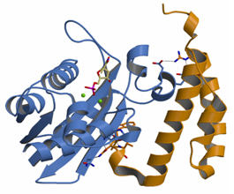 Figure 2: Crystal structure of the exonuclease epsilon subunit of DNA polymerase III in complex with the HOT protein. From this structure one can infer the DNA polymerase III epsilon:theta complex (Kirby et al, J Biol Chem, 281:38466-71 (2006).
|
|