|
Methamphetamine-commonly known as "speed," "meth," "ice," or "crystal"-is a powerfully addictive stimulant that acts on the central nervous system to produce increased wakefulness and physical activity as well as irritability, insomnia, confusion, tremors, convulsions, anxiety, paranoia, and aggressiveness. The drug increases heart rate and blood pressure and can irreversibly damage blood vessels in the brain. Now, NIDA-supported research has demonstrated that methamphetamine abusers risk long-term brain damage.
Dr. Thomas Ernst and Dr. Linda Chang at the Harbor-UCLA Medical Center in Torrance, California, used a noninvasive brain imaging technique called magnetic resonance spectroscopy (MRS) to measure levels of brain chemicals that indicate whether brain cells are healthy or are diseased or damaged. "We found abnormal brain chemistry in methamphetamine users in all the brain regions we studied. In one of the regions, the amount of damage was also related to the history of drug use-those abusers who had the greatest cumulative lifetime methamphetamine use had the strongest indications of cell damage," Dr. Chang says.
In each of the participants, the researchers examined a midfrontal region consisting largely of "gray matter"-nerve cell bodies and short extensions called dendrites that communicate with neighboring neurons-and an area in the basal ganglia, a neuron-dense region at the top of the brain stem. They also examined a right frontal area composed largely of "white matter," or long nerve cell extensions called axons that communicate with more distant regions of the brain. These brain regions were selected because they are areas of high activity of dopamine, a neurotransmitter involved in the "rush" and pleasure associated with addictive drugs.
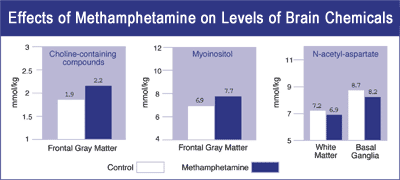 Neurons in three areas of the brain show changes in levels of brain chemicals that serve as indicators of health of brain cells. Levels of choline-containing compounds and myo-inositol are elevated and levels of N-acetyl-aspartate are reduced in methamphetamine abusers who have not used the drug for at least 2 weeks and up to 21 months. The changes in concentrations of these chemical markers suggest that methamphetamine use may result in long-term damage to brain cells used in thinking. |
The researchers measured levels of N-acetyl-aspartate (NAA), a metabolite produced only in neurons. "NAA levels are a measure of the viability and density of neurons," Dr. Ernst says. "Many diseases associated with brain cell loss or damage, such as Alzheimer's disease, stroke, and epilepsy, are also associated with reduced NAA."
The scientists also measured levels of two other chemical markers-choline-containing compounds and myo-inositol (MI)-associated mostly with specialized cells called glial cells. "The primary role of glial cells is to maintain normal function of neurons, including repair of injury to the cells. Increases in glial markers suggest proliferation of these support cells in response to neuronal damage," Dr. Ernst explains.
Dr. Chang and Dr. Ernst measured levels of the chemical markers in 26 participants who had a history of methamphetamine abuse but had not used the drug for periods ranging from 2 weeks to 21 months with a median of 4.25 months since last use. They compared the results to measurements of 24 participants who had no history of methamphetamine use.
Among the methamphetamine abusers, NAA levels were reduced by 5 percent in the frontal white matter and 6 percent in the basal ganglia compared with levels among nonusers. "The reduced concentrations of NAA in the drug users' brains suggest that long-term methamphetamine abuse results in loss of or damage to neurons," Dr. Ernst says. Methamphetamine abusers also showed increases of 11 percent in levels of MI and 13 percent in levels of choline-containing compounds in the frontal gray matter compared with nonusers. "This suggests an increased number or size of glial cells as a reaction to the injurious effects of the drug," he adds.
"Methamphetamine may be substantially toxic to the cells we use in thinking," Dr. Ernst says. "This long-term, and perhaps permanent, alteration in basic brain chemistry is additional evidence that methamphetamine abuse, like abuse of other drugs, should be considered a brain disease and treated accordingly."
Sources
Ernst, T.; Chang, L.; Leonido-Lee, M.; and Speck, O. Evidence for long-term neurotoxicity associated with methamphetamine abuse: a 1H MRS study. Neurology 54(6):1344-1349, 2000.
|