|
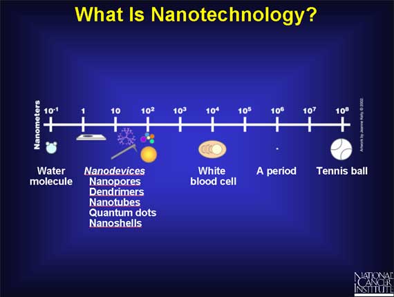
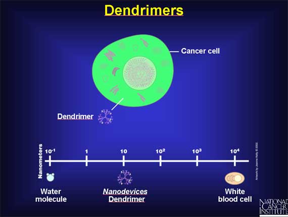
A nanometer is a billionth of a meter. It's difficult to imagine anything so small, but think of something only 1/80,000 the width of a human hair. Ten hydrogen atoms could be laid side-by-side in a single nanometer.
Nanotechnology is the creation of useful materials, devices, and systems through the manipulation of matter on this miniscule scale. The emerging field of nanotechnology involves scientists from many different disciplines, including physicists, chemists, engineers, and biologists.
There are many interesting nanodevices being developed that have a potential to improve cancer detection, diagnosis, and treatment.
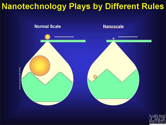
Much of today's nanoscale research is designed to reach a better understanding of how matter behaves on this small scale. The factors that govern larger systems do not necessarily apply at the nanoscale. Because nanomaterials have large surface areas relative to their volumes, phenomena like friction and sticking are more important than they are in larger systems.
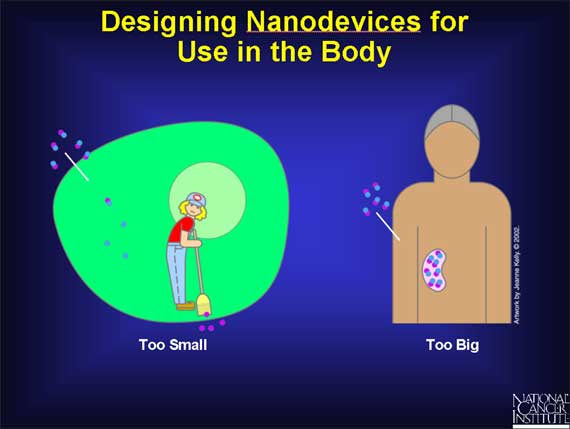
Other challenges apply specifically to the use of nanostructures within biological systems. Nanostructures can be so small that the body may clear them too rapidly for them to be effective in detection or imaging. Larger nanoparticles may accumulate in vital organs, creating a toxicity problem. Scientists will need to consider these factors as they attempt to create nanodevices the body will accept.
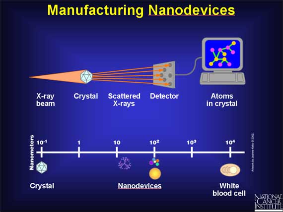
There are two basic approaches for creating nanodevices. Scientists refer to these methods as the top-down approach and the bottom-up approach. The top-down approach involves molding or etching materials into smaller components. This approach has traditionally been used in making parts for computers and electronics. The bottom-up approach involves assembling structures atom-by-atom or molecule-by-molecule, and may prove useful in manufacturing devices used in medicine.
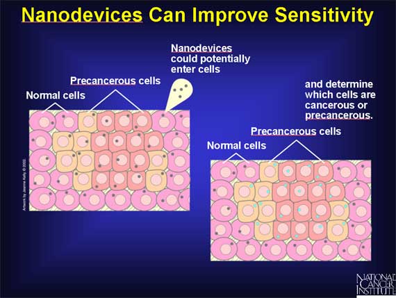
Most animal cells are 10,000 to 20,000 nanometers in diameter. This means that nanoscale devices (less than 100 nanometers) can enter cells and the organelles inside them to interact with DNA and proteins. Tools developed through nanotechnology may be able to detect disease in a very small amount of cells or tissue. They may also be able to enter and monitor cells within a living body.

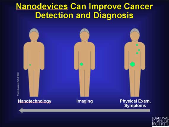
Detection of cancer at early stages is a critical step in improving cancer treatment. Currently, detection and diagnosis of cancer usually depend on changes in cells and tissues that are detected by a doctor's physical touch or imaging expertise. Instead, scientists would like to make it possible to detect the earliest molecular changes, long before a physical exam or imaging technology is effective. To do this, they need a new set of tools.
In order to successfully detect cancer at its earliest stages, scientists must be able to detect molecular changes even when they occur only in a small percentage of cells. This means the necessary tools must be extremely sensitive. The potential for nanostructures to enter and analyze single cells suggests they could meet this need.
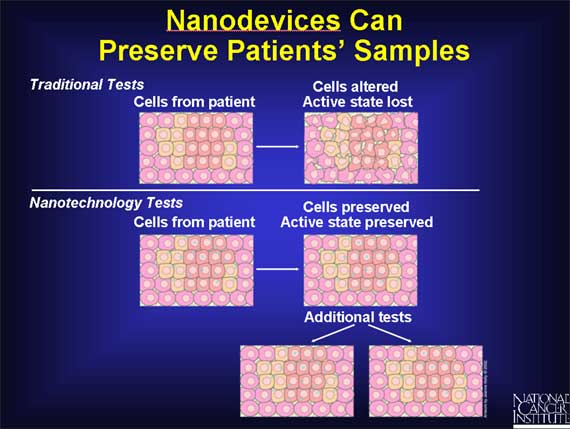
Many nanotechnology tools will make it possible for clinicians to run tests without physically altering the cells or tissue they take from a patient. This is important because the samples clinicians use to screen for cancer are often in limited supply. It is also important because it can capture and preserve cells in their active state. Scientists would like to perform tests without altering cells, so the cells can be used again if further tests are needed.
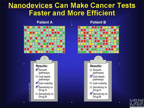
Miniaturization will allow the tools for many different tests to be situated together on the same small device. Researchers hope that nanotechnology will allow them to run many diagnostic tests simultaneously.
One nanodevice that can improve cancer detection and diagnosis is the cantilever. These tiny levers, which are anchored at one end, can be engineered to bind to molecules that represent some of the changes associated with cancer. They may bind to altered DNA sequences or proteins that are present in certain types of cancer. When these molecules bind to the cantilevers, surface tension changes, causing the cantilevers to bend. By monitoring the bending of the cantilevers, scientists can tell whether molecules are present. Scientists hope this property will prove effective when cancer-associated molecules are present--even in very low concentrations--making cantilevers a potential tool for detecting cancer in its early stages.

 |
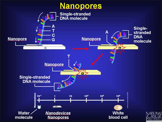
Another interesting nanodevice is the nanopore. Improved methods of reading the genetic code will help researchers detect errors in genes that may contribute to cancer. Scientists believe nanopores, tiny holes that allow DNA to pass through one strand at a time, will make DNA sequencing more efficient. As DNA passes through a nanopore, scientists can monitor the shape and electrical properties of each base, or letter, on the strand. Because these properties are unique for each of the four bases that make up the genetic code, scientists can use the passage of DNA through a nanopore to decipher the encoded information, including errors in the code known to be associated with cancer.
Another nanodevice that will help identify DNA changes associated with cancer is the nanotube. Nanotubes are carbon rods about half the diameter of a molecule of DNA that not only can detect the presence of altered genes, but they may help researchers pinpoint the exact location of those changes.
To prepare DNA for nanotube analysis, scientists must attach a bulky molecule to regions of the DNA that are associated with cancer. They can design tags that seek out specific mutations in the DNA and bind to them.
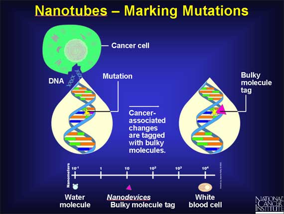
Once the mutation has been tagged, researchers use a nanotube tip resembling the needle on a record player to trace the physical shape of DNA and pinpoint the mutated regions. The nanotube creates a map showing the shape of the DNA molecule, including the tags identifying important mutations. Since the location of mutations can influence the effects they have on a cell, these techniques will be important in predicting disease.

|
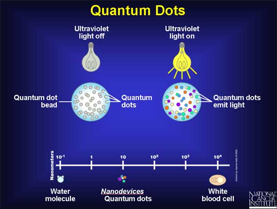
Another minuscule molecule that will be used to detect cancer is a quantum dot. Quantum dots are tiny crystals that glow when they are stimulated by ultraviolet light. The wavelength, or color, of the light depends on the size of the crystal. Latex beads filled with these crystals can be designed to bind to specific DNA sequences. By combining different sized quantum dots within a single bead, scientists can create probes that release distinct colors and intensities of light. When the crystals are stimulated by UV light, each bead emits light that serves as a sort of spectral bar code, identifying a particular region of DNA.
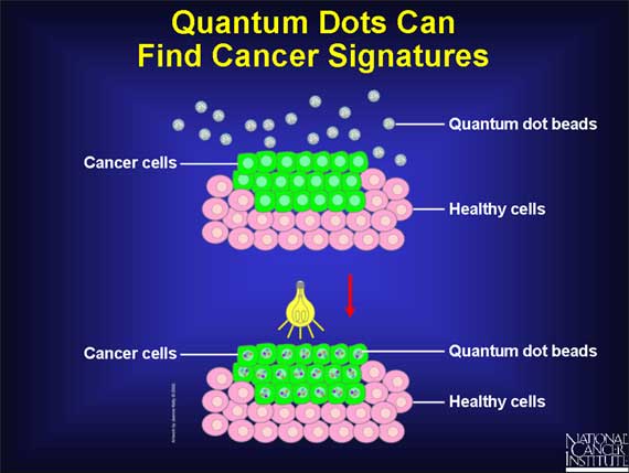
To detect cancer, scientists can design quantum dots that bind to sequences of DNA that are associated with the disease. When the quantum dots are stimulated with light, they emit their unique bar codes, or labels, making the critical, cancer-associated DNA sequences visible.
The diversity of quantum dots will allow scientists to create many unique labels, which can identify numerous regions of DNA simultaneously. This will be important in the detection of cancer, which results from the accumulation of many different changes within a cell.
Another advantage of quantum dots is that they can be used in the body, eliminating the need for biopsy.
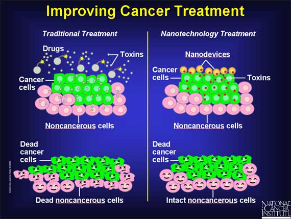
Nanotechnology may also be useful for developing ways to eradicate cancer cells without harming healthy, neighboring cells. Scientists hope to use nanotechnology to create therapeutic agents that target specific cells and deliver their toxin in a controlled, time-released manner.
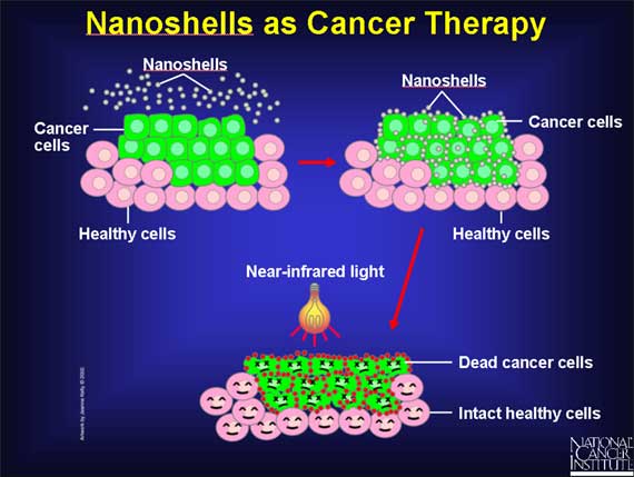
Nanoshells are miniscule beads coated with gold. By manipulating the thickness of the layers making up the nanoshells, scientists can design these beads to absorb specific wavelengths of light. The most useful nanoshells are those that absorb near-infrared light, which can easily penetrate several centimeters of human tissue. The absorption of light by the nanoshells creates an intense heat that is lethal to cells.

Researchers can already link nanoshells to antibodies that recognize cancer cells. Scientists envision letting these nanoshells seek out their cancerous targets, then applying near-infrared light. In laboratory cultures, the heat generated by the light-absorbing nanoshells has successfully killed tumor cells while leaving neighboring cells intact.
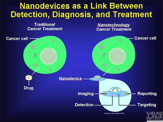
Researchers aim eventually to create nanodevices that do much more than deliver treatment. The goal is to create a single nanodevice that will do many things: assist in imaging inside the body, recognize precancerous or cancerous cells, release a drug that targets only those cells, and report back on the effectiveness of the treatment.
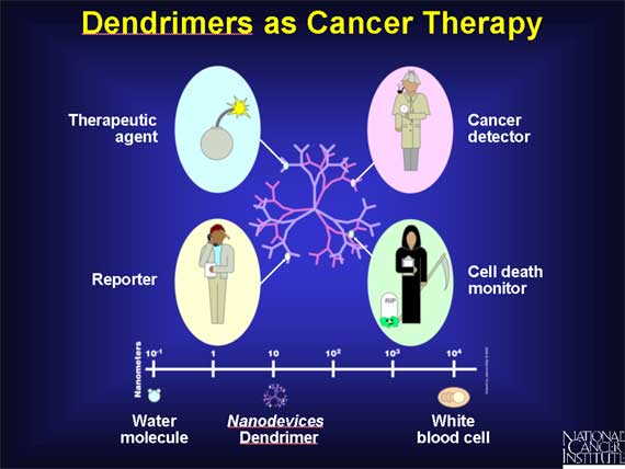
Research is being done on a number of nanoparticles created to facilitate drug delivery. One such molecule with potential to link treatment with detection and diagnosis is known as a dendrimer. Dendrimers are man-made molecules about the size of an average protein, and have a branching shape. This shape gives them vast amounts of surface area to which scientists can attach therapeutic agents or other biologically active molecules. Researchers aim eventually to create nanodevices that do much more than deliver treatment.
A single dendrimer can carry a molecule that recognizes cancer cells, a therapeutic agent to kill those cells, and a molecule that recognizes the signals of cell death. Researchers hope to manipulate dendrimers to release their contents only in the presence of certain trigger molecules associated with cancer. Following drug release, the dendrimers may also report back whether they are successfully killing their targets.
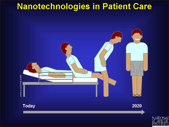
Nanotechnologies that will aid in cancer care are in various stages of discovery and development. Experts believe that quantum dots, nanopores, and other devices for detection and diagnosis may be available for clinical use in 5 to 15 years. Therapeutic agents are expected to be available within a similar time frame. Devices that integrate detection and therapy could be used clinically in about 15 or 20 years.
|


