How the Heart Works
Your child’s heart is a muscle about the size of his or her fist. It works like a pump and beats 100,000 times a day.
The heart has two sides, separated by an inner wall called the septum. The right side of the heart pumps blood to the lungs to pick up oxygen. Then, oxygen-rich blood returns from the lungs to the left side of the heart, and the left side pumps it to the body.
The heart has four chambers and four valves and is connected to various blood vessels. Veins are the blood vessels that carry blood from the body to the heart. Arteries are the blood vessels that carry blood away from the heart to the body.
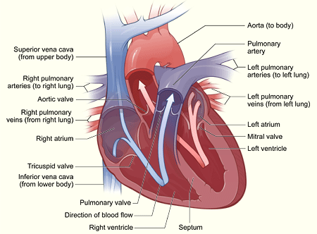
A Healthy Heart Cross-Section
The illustration shows a cross-section of a healthy heart and its inside structures. The blue arrow shows the direction in which oxygen-poor blood flows from the body to the lungs. The red arrow shows the direction in which oxygen-rich blood flows from the lungs to the rest of the body.
Heart Chambers
The heart has four chambers or "rooms."
- The atria (AY-tree-uh) are the two upper chambers that collect blood as it comes into the heart.
- The ventricles (VEN-trih-kuls) are the two lower chambers that pump blood out of the heart to the lungs or other parts of the body.
Heart Valves
Four valves control the flow of blood from the atria to the ventricles and from the ventricles into the two large arteries connected to the heart.
- The tricuspid (tri-CUSS-pid) valve is in the right side of the heart, between the right atrium and the right ventricle.
- The pulmonary (PULL-mun-ary) valve is in the right side of the heart, between the right ventricle and the entrance to the pulmonary artery, which carries blood to the lungs.
- The mitral (MI-trul) valve is in the left side of the heart, between the left atrium and the left ventricle.
- The aortic (ay-OR-tik) valve is in the left side of the heart, between the left ventricle and the entrance to the aorta, the artery that carries blood to the body.
Valves are like doors that open and close. They open to allow blood to flow through to the next chamber or to one of the arteries, and then they shut to keep blood from flowing backward.
When the heart's valves open and close, they make a "lub-DUB" sound that a doctor can hear using a stethoscope.
- The first sound—the “lub”—is made by the mitral and tricuspid valves closing at the beginning of systole (SIS-toe-lee). Systole is when the ventricles contract, or squeeze, and pump blood out of the heart.
- The second sound—the “DUB”—is made by the aortic and pulmonary valves closing at beginning of diastole (di-AS-toe-lee). Diastole is when the ventricles relax and fill with blood pumped into them by the atria.
Arteries
The arteries are major blood vessels connected to your heart.
- The pulmonary artery carries blood pumped from the right side of the heart to the lungs to pick up a fresh supply of oxygen.
- The aorta is the main artery that carries oxygen-rich blood pumped from the left side of the heart out to the body.
- The coronary arteries are the other important arteries attached to the heart. They carry oxygen-rich blood from the aorta to the heart muscle, which must have its own blood supply to function.
Veins
The veins are also major blood vessels connected to your heart.
- The pulmonary veins carry oxygen-rich blood from the lungs to the left side of the heart so it can be pumped out to the body.
- The vena cava is a large vein that carries oxygen-poor blood from the body back to the heart.
For more information on how a healthy heart works, see the Diseases and Conditions Index article on How the Heart Works. This article contains animations that show how your heart pumps blood and how your heart’s electrical system works.
The Heart With Patent Ductus Arteriosus
The ductus arteriosus is a blood vessel that
connects a baby's aorta and pulmonary artery while the baby is in the womb.
This connection allows blood to be pumped from the right side of the heart
straight to the aorta without stopping at the lungs for oxygen. In the womb,
only a small amount of a baby's blood needs to go to the lungs because the baby
gets oxygen from the mother's bloodstream. The baby's pulmonary artery, which
carries blood to the lungs, is not needed at this time.
After birth, the baby is no longer connected to the
mother's bloodstream. The baby's blood must now go to his or her own lungs to
get oxygen. Normally, as the baby begins to breathe on his or her own, the
pulmonary artery opens to allow blood into the lungs, and the ductus arteriosus
closes. Once the ductus arteriosus closes, blood leaving the right side of the
heart no longer goes straight to the aorta. First, it goes through the
pulmonary arteries and stops at the lungs to pick up oxygen. Then, blood
carrying oxygen returns to the left side of the heart and is pumped out to the
rest of the body.
If the ductus arteriosus does not close after birth
as it should, it is called a patent ductus arteriosus (PDA). A PDA allows blood
to flow directly from the aorta into the pulmonary artery and from there to the
lungs. This extra amount of blood flowing into the lungs strains the heart and
increases the blood pressure in the arteries of the lungs.
Effects of patent ductus arteriosus
Normal birth-weight infants. The
larger a PDA is, the greater the amount of extra blood that passes through the
lungs. A small PDA might not cause any problems, whereas a larger PDA is likely
to cause problems.
PDA can increase the risk of
bacterial endocarditis. Bacterial endocarditis is an infection
of the lining of the heart, valves, or arteries. In the case of PDA, the
increased flow of blood can irritate the lining of the pulmonary artery where
the PDA connects. This irritation makes it easier for bacteria in the
bloodstream to collect and grow there.
A large PDA that is allowed to remain open for an
extended period of time can cause the heart to enlarge and to have to work
harder. Also, fluid can build up in the lungs.
Premature infants. For premature
infants (babies born too early), PDA can be more serious than in normal-weight
babies. Preemies with PDA are more likely to have damage to their lungs from
the extra blood flowing through the PDA. Preemies with PDA may need to be on a
ventilator to help them with their breathing.
The increased flow of blood through the lungs also
can reduce blood flow to the rest of the body. This can damage other organs,
especially the intestines and kidneys. |